
Tarsometatarsal joints
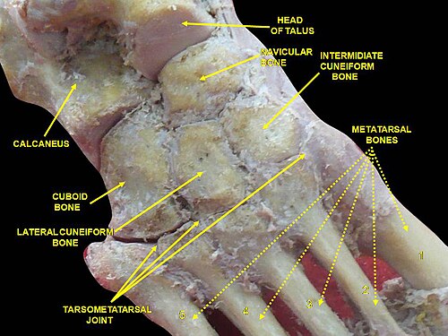
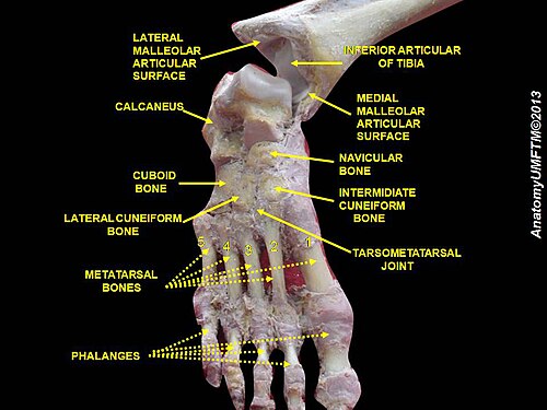
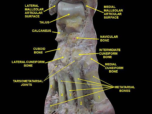
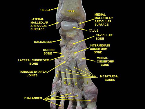
The tarsometatarsal joints (Lisfranc joints) are arthrodial joints in the foot. The tarsometatarsal joints involve the first, second and third cuneiform bones, the cuboid bone and the metatarsal bones. The eponym of Lisfranc joint is 18th–19th-century surgeon and gynecologistJacques Lisfranc de St. Martin.[1]
Structure
Bones
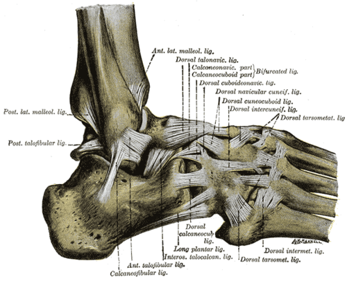
The bones entering into their formation are the first, second, and third cuneiforms, and the cuboid bone, which articulate with the bases of the metatarsal bones.
The first metatarsal bone articulates with the first cuneiform; the second is deeply wedged in between the first and third cuneiforms articulating by its base with the second cuneiform; the third articulates with the third cuneiform; the fourth, with the cuboid and third cuneiform; and the fifth, with the cuboid.
The bones are connected by dorsal, plantar, and interosseous ligaments.
Dorsal ligaments
The dorsal ligaments are strong, flat bands.
The first metatarsal is joined to the first cuneiform by a broad, thin band; the second has three, one from each cuneiform bone; the third has one from the third cuneiform; the fourth has one from the third cuneiform and one from the cuboid; and the fifth, one from the cuboid.
Plantar ligaments
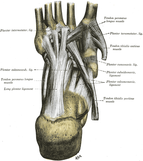
The plantar ligaments consist of longitudinal and oblique bands, disposed with less regularity than the dorsal ligaments.
Those for the first and second metatarsals are the strongest; the second and third metatarsals are joined by oblique bands to the first cuneiform; the fourth and fifth metatarsals are connected by a few fibers to the cuboid.
Interosseous ligaments
The interosseous ligaments are three in number.
- The first is the strongest, and passes from the lateral surface of the first cuneiform to the adjacent angle of the second metatarsal.
- The second connects the third cuneiform with the adjacent angle of the second metatarsal.
- The third connects the lateral angle of the third cuneiform with the adjacent side of the base of the third metatarsal.
Synovial membrane
The synovial membrane between the first cuneiform and the first metatarsal forms a distinct sac.
الغشاء الزلالي بين العظمين الإسفينيين الثاني والثالث من الخلف، والعظمين المشطيين الثاني والثالث من الأمام، هو جزء من الغشاء الزلالي الرسغي الكبير .
يتم إرسال امتدادين للأمام منه، أحدهما بين الجانبين المتجاورين للعظمين الثاني والثالث، والآخر بين عظام مشط القدم الثالث والرابع.
يشكل الغشاء الزلالي بين العظم المكعبي وعظام مشط القدم الرابعة والخامسة كيساً مميزاً.
ومنه يمتد امتداد إلى الأمام بين عظمي مشط القدم الرابع والخامس.
وظيفة
تقتصر الحركات المسموح بها بين عظام الرسغ وعظام مشط القدم على انزلاق طفيف للعظام فوق بعضها البعض.
الأهمية السريرية
تُعد إصابة ليزفرانك شائعة بين الرياضيين.
مراجع
 تتضمن هذه المقالة نصًا في الملكية العامة من الصفحة 358 من الطبعة العشرين من كتاب تشريح غراي (1918).
روابط خارجية
صور إضافية
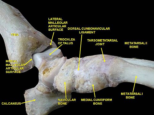 مفصل الكاحل. تشريح عميق.
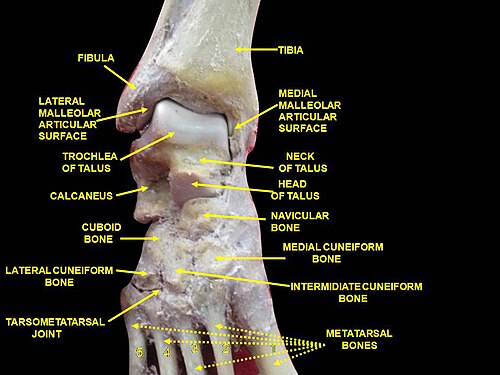 مفصل الكاحل. تشريح عميق.
 مفصل الكاحل. تشريح عميق.
 مفصل الكاحل. تشريح عميق.
 مفاصل الكاحل والرسغ. عظام القدم. تشريح عميق.
 مفاصل الكاحل والرسغ. عظام القدم. تشريح عميق.
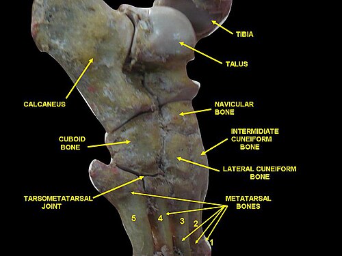 مفصل الكاحل. عظام القدم. تشريح عميق.
- مفاصل الطرف السفلي