
Allergic contact dermatitis
Allergic contact dermatitis (ACD) is a form of contact dermatitis that is the manifestation of a type IV hypersensitivity (delayed hypersensitivity) response caused by contact with a substance; the other type being irritant contact dermatitis (ICD). It differs from classical allergic reactions, which are type I hypersensitivity reactions.[1]
Although less common than ICD, ACD is accepted to be the most prevalent form of immunotoxicity found in humans.[2] By its hypersensitive nature, this form of contact dermatitis is atypical within the population. The mechanisms by which these reactions occur are complex, with many levels of fine control. Their immunology centres on the interaction of immunoregulatory cytokines and discrete subpopulations of T lymphocytes.
Signs and symptoms
The symptoms of allergic contact dermatitis are very similar to those caused by irritant contact dermatitis, which makes the first even harder to diagnose. The first sign of allergic contact dermatitis is the presence of a rash or skin lesion at the site of exposure.[3] Depending on the type of allergen causing it, the rash can ooze, drain, or crust, and it can become raw, scaled, or thickened. Also, it is possible that the skin lesion does not take the form of a rash, but it may include papules, blisters, vesicles, or even a simple red area. The main difference between the rash caused by allergic contact dermatitis and the one caused by irritant contact dermatitis is that the latter tends to be confined to the area where the trigger touched the skin, whereas in allergic contact dermatitis, the rash is more likely to be more widespread on the skin.[4] Another characteristic of the allergic contact dermatitis rash is that it usually appears after a day or two after exposure to the allergen, unlike irritant contact dermatitis that appears immediately after the contact with the trigger.
Other symptoms may include itching, skin redness or inflammation, localized swelling, and the area may become more tender or warmer. If left untreated, the skin may darken and become leathery and cracked.[5] Pain can also be present. Dermatitis can occur anywhere on the skin, but is most common on the hands (22% of people), scattered across the body (18%), or on the face (17%).[6]
The rash and other symptoms typically occur 24 to 48 hours after the exposure; in some cases, the rash may persist for weeks.[3] Once an individual has developed a skin reaction to a certain substance it is most likely that they will have it for the rest of their life, and the symptoms will reappear when in contact with the allergen.
Cause
Common allergens implicated include the following:
- Bacitracin – topical antibiotic found by itself, or as Polysporin or Triple Antibiotic
- Balsam of Peru (Myroxylon pereirae) – used in food and drink for flavoring, in perfumes and toiletries for fragrance, and in medicine and pharmaceutical items for healing properties; derived from tree resin.[7][8][9] It may also be a component of artificial vanilla and/or cinnamon flavorings.[7]
- Chromium – used in the tanning of leather. Also a component of uncured cement/mortar, facial cosmetics and some bar soaps.
- Cobalt chloride – metal found in medical products; hair dye; antiperspirant; metal-plated objects such as snaps, buttons or tools; and in cobalt blue pigment
- Colophony (Rosin) – rosin, sap, or sawdust typically from spruce or fir trees
- Formaldehyde – preservative with multiple uses, e.g., in paper products, paints, medications, household cleaners, cosmetic products, and fabric finishes. Often released into products by the use of formaldehyde releasers such as imidazolidinyl urea, diazolidinyl urea, Quaternium-15, DMDM Hydantoin, and 2-bromo-2-nitropropane-1,3-diol.
- Fragrance mix – group of the eight most common fragrance allergens found in foods, cosmetic products, insecticides, antiseptics, soaps, perfumes, and dental products [10] Limonene and Linalool are also commonly implicated in allergic contact dermatitis responsible for over 10% of all cases of Allergic contact dermatitis[11]
- Gold (gold sodium thiosulfate) – precious metal and compound often found in jewelry and dental materials
- Isothiazolinones – preservatives used in many personal care, household, and commercial products.
- Mercaptobenzothiazole – in rubber products, notably shoes, gloves, and car tires.
- Neomycin – topical antibiotic common in first aid creams and ointments, cosmetics, deodorant, soap, and pet food. Found by itself, or in Neosporin or Triple Antibiotic
- Nickel (nickel sulfate hexahydrate) – has been recognized as a significant cause of allergy.[12] This metal is frequently encountered in stainless steel cookware,[13] jewelry and clasps or buttons on clothing. Current estimates gauge are that roughly 2.5 million US adults and 250,000 children suffer from nickel allergy, which costs an estimated $5.7 billion per year for treatment of symptoms.[14] A significant portion of nickel allergy is preventable.[15]
- p-Phenylenediamine (PPD) – although it's mainly used as a component of engineering polymers and composites like kevlar, it is also an ingredient in hair dyes, which, once sensitized to, becomes lifelong. One can develop active sensitization to products, including but not limited to black clothing, various inks, hair dye, dyed fur, dyed leather, and certain photographic products.
- Photographic developers, especially those containing metol
- Quaternium-15 – preservative in cosmetic products (self-tanners, shampoo, nail polish, sunscreen) and in industrial products (polishes, paints, and waxes).[16]
- Sap from certain species of mangrove and agave
- Soluble salts of platinum – seeplatinosis
- Thiomersal – mercury compound used in local antiseptics and in vaccines
- Topical anesthetics – such as pramoxine or diphenhydramine, after prolonged use
- Topical steroid – seesteroid allergy
- Urushiol – oily coating from plants of Toxicodendron genus – poison ivy, poison oak, and poison sumac. Also found in mango plants, mango skin, cashews, and smoke from burning urushiol-containing plants, which can cause skin as well as severe lung irritation.
Mechanism
ينشأ التهاب الجلد التماسي التحسسي (ACD) نتيجة مرحلتين أساسيتين: مرحلة التحريض ، التي تُهيئ الجهاز المناعي وتُحسّسه للاستجابة التحسسية، ومرحلة الاستثارة ، التي تُحفز فيها هذه الاستجابة. [ 2 ] خلال مرحلة التحريض، يؤدي التعرض للمادة المسببة للحساسية ( المؤرج ) إلى معالجتها وعرضها بواسطة الخلايا المناعية، مما يُهيئ الجهاز المناعي للاستجابة. في مرحلة الاستثارة، عند التلامس اللاحق مع نفس المؤرج، تتعرف الخلايا التائية على المؤرج مباشرةً، مما يؤدي إلى استجابة مناعية في موضع التلامس. يُصنف رد الفعل التحسسي، الذي تتوسطه الخلايا التائية مباشرةً، على أنه تفاعل فرط حساسية من النوع الرابع. يختلف هذا التصنيف عن فرط الحساسية من النوع الأول الأكثر شيوعًا (مثل حمى القش )، حيث ترتبط المؤرجات بالأجسام المضادة IgE، التي بدورها تُحفز الخلايا البدينة .
في حالات الحساسية التلامسية، تكون الجزيئات المسؤولة (مسببات الحساسية) صغيرة الحجم عادةً، ولا يستطيع الجهاز المناعي التعرف عليها مباشرةً. لا تُثير هذه المسببات رد فعل تحسسي إلا بعد خضوعها لعملية تُسمى التَحَوُّل . خلال هذه العملية، ترتبط مسببات الحساسية بجزيئات أكبر (بروتينات حاملة) موجودة بشكل طبيعي في الجلد. هذا المركب من مسبب الحساسية والبروتين الحامل هو ما يتعرف عليه الجهاز المناعي كجسم غريب، مما يؤدي إلى رد فعل تحسسي.
تُعتبر الخلايا اللانغرهانسية (LCs) (وفي بعض الحالات الخلايا المتغصنة الأخرى (DCs)) المركب الناتج جسمًا غريبًا، فتقوم هذه الخلايا بابتلاع البروتين ونقله عبر الجهاز اللمفاوي إلى العقد اللمفاوية الإقليمية ، ثم تعرض المستضد للخلايا اللمفاوية التائية . تُسيطر السيتوكينات والكيموكينات على هذه العملية ، ومنها عامل نخر الورم ألفا (TNF-α) وبعض أفراد عائلة الإنترلوكين (1، 13، و18)، حيث يعمل تأثيرها إما على تعزيز أو تثبيط حركة وهجرة هذه الخلايا اللانغرهانسية. [ 2 ] وأثناء انتقال الخلايا اللانغرهانسية إلى العقد اللمفاوية، تتمايز وتتحول إلى خلايا متغصنة، وهي خلايا محفزة للمناعة.
بمجرد دخولها إلى الغدد اللمفاوية ، تقوم الخلايا المتغصنة المتمايزة بعرض الجزء المستضدي المرتبط بمسبب الحساسية على الخلايا اللمفاوية التائية. ثم تنقسم هذه الخلايا التائية وتتمايز، وتتكاثر استنساخياً بحيث إذا تعرض الشخص لمسبب الحساسية مرة أخرى، فإن هذه الخلايا التائية ستستجيب بشكل أسرع وأكثر شراسة.
أشار وايت وآخرون إلى وجود عتبة لآليات التحسس التحسسي الناتج عن مسببات الحساسية المرتبطة بالتهاب الجلد التماسي التحسسي (1986). [ 17 ] يُعتقد أن هذه العتبة مرتبطة بالمستوى الذي يحفز عنده السم زيادة إفراز السيتوكينات والكيموكينات الضرورية. كما اقتُرح أن الوسيلة التي يصل بها مسبب الحساسية إلى الجلد قد تُسهم في تحسس البشرة، وذلك من خلال تسهيل اختراق الجلد وإحداث نوع من الصدمة وتحفيز إفراز السيتوكينات.
استجابة الذاكرة
بمجرد أن يُصاب الشخص بحساسية تجاه مادة مُسببة للحساسية، فإن أي تلامس لاحق معها قد يُثير رد فعل تحسسي، يُعرف عادةً باسم استجابة الذاكرة، في موضع التحسس الأصلي. فعلى سبيل المثال، إذا كان الشخص يُعاني من التهاب الجلد التماسي التحسسي في الجفون، نتيجة استخدام مستحضرات التجميل مثلاً، فإن لمس المادة المُسببة للحساسية بالأصابع قد يُثير رد فعل تحسسي في الجفون.
يعود ذلك إلى خلايا الذاكرة التائية الجلدية الموضعية، التي تبقى في موقع التحسس الأصلي. وبالمثل، تقوم الخلايا اللمفاوية التائية السامة بدوريات في منطقة من الجلد، وتلعب دورًا هامًا في السيطرة على إعادة تنشيط الفيروسات (مثل فيروس الهربس البسيط ) والحد من تكاثرها عند إعادة تنشيطها. [ 18 ] عادةً ما تستغرق استجابة الذاكرة، أو "تفاعل إعادة الاختبار"، من يومين إلى ثلاثة أيام بعد التعرض لمسبب الحساسية، وقد تستمر لمدة تتراوح بين أسبوعين وأربعة أسابيع.
تشخبص
يعتمد تشخيص التهاب الجلد التماسي التحسسي بشكل أساسي على الفحص السريري والتاريخ الطبي . في بعض الحالات، يستطيع الأطباء وضع تشخيص دقيق بناءً على الأعراض التي يعاني منها المريض ومظهر الطفح الجلدي. في حالة الإصابة بنوبة واحدة من التهاب الجلد التماسي التحسسي، يكون هذا كافيًا. أما الطفح الجلدي المزمن أو المتقطع الذي لا يمكن تفسيره بسهولة من خلال التاريخ الطبي والفحص السريري، فغالبًا ما يحتاج إلى مزيد من الفحوصات.
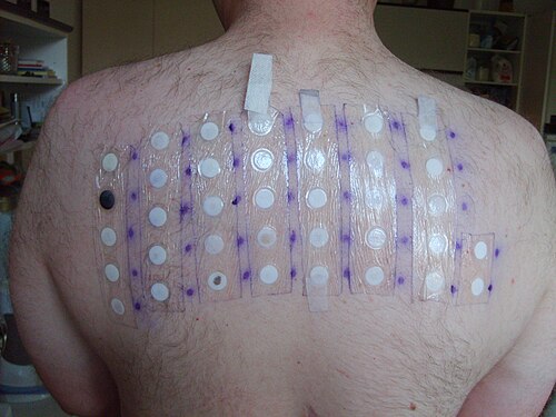
يُعد اختبار الرقعة (اختبار فرط الحساسية المتأخر بالتلامس) [ 19 ] فحصًا شائع الاستخدام لتحديد السبب الدقيق لالتهاب الجلد التماسي التحسسي. ووفقًا للأكاديمية الأمريكية للحساسية والربو والمناعة، فإن "اختبار الرقعة هو المعيار الذهبي لتحديد مسببات الحساسية بالتلامس". [ 3 ]
يتضمن اختبار الرقعة وضع كميات صغيرة من المواد المسببة للحساسية المحتملة على رقع صغيرة، ثم توضع هذه الرقع على الجلد. [ 20 ] بعد يومين، تُزال الرقع، وإذا حدث رد فعل تحسسي تجاه إحدى المواد المستخدمة، فسيظهر نتوء بارز أسفل الرقعة. تُعاد قراءة نتائج الاختبار بعد 72 أو 96 ساعة من وضع الرقعة.
Patch testing is used for patients who have chronic, recurring contact dermatitis.[3] Other tests that may be used to diagnose contact dermatitis and rule out other potential causes of the symptoms include a skin biopsy and culture of the skin lesion.
Treatment
The clinical expression of the dermatitis can be mitigated by avoidance of the allergen. Through compliance with avoidance measures, the immune system can become less stimulated. The key to avoidance is proper evaluation and detection of the inciting allergen. However, once the immune system registers the allergen, the recognition is usually permanent.
The first step in treating the condition is appropriate recognition of the clinical problem, followed by identification of the culprit chemical and the source of that chemical. Corticosteroid creams should be used carefully and according to the prescribed directions because, when overused over longer periods of time, they can cause thinning of the skin. Also, in some instances, such as poison ivy dermatitis calamine lotion and cool oatmeal baths may relieve itching.[21]
Unlike the more common Type I allergies (e.g., hay fever), contact allergies are not mediated by histamine, making the use of normal allergy medication (antihistamine) medication ineffective, and other drugs have to be used to treat the allergic reaction.
Usually, severe cases are treated with systemic corticosteroids, which may be tapered gradually, with various dosing schedules ranging from a total of 12 – 20 days to prevent the recurrence of the rash (while the chemical allergen is still in the skin, up to 3 weeks, as well as a topical corticosteroid.[3] Tacrolimus ointment or pimecrolimus cream can also be used additionally to the corticosteroid creams or instead of these. Oral antihistamines such as diphenhydramine or hydroxyzine may also be used in more severe cases to relieve the intense itching via sedation.[22] Topical antihistamines are not advised as there might be a second skin reaction (treatment associated contact dermatitis) from the lotion itself.
The other symptoms caused by allergic contact dermatitis may be eased with cool compresses to stop the itching. It is vital for treatment success that the trigger be identified and avoided. The discomfort caused by the symptoms may be relieved by wearing smooth-textured cotton clothing to avoid frictional skin irritation or by avoiding soaps with perfumes and dyes. Commonly, the symptoms may resolve without treatment in 2 to 4 weeks, but specific medication may hasten the healing as long as the trigger is avoided. Also, the condition might become chronic if the allergen is not detected and avoided.
Identification of the allergen can be aided by the site of the dermatitis. Allergic dermatitis of the hands is often due to contact with preservatives, fragrances, metals, rubber, or topical antibiotics.[6] Dermatitis at the front of the face is often due to gold (from jewelry and foundation), make-up, moisturizers, wrinkle creams, and topical medication.[6] Along the eyelids as well as the sides of the head and neck, dermatitis is often caused by shampoo and conditioner dripping down from the hair.[6] Inflammation on one side of the face often suggests transfer of an allergen from the hands or from the face of a partner.[6]
Epidemiology
Allergic contact dermatitis is common, affecting up to 20% of all people.[6] People sensitive to one allergen are at an increased risk of being sensitive to others.[6] Family members of those with allergic contact dermatitis are at higher risk of developing it themselves.[6] Women are at higher risk of developing allergic contact dermatitis than men.[6]
See also
References
- ↑Martín A, Gallino N, Gagliardi J, Ortiz S, Lascano AR, Diller A, Daraio MC, Kahn A, Mariani AL, Serra HM (August 2002). "Early inflammatory markers in elicitation of allergic contact dermatitis". BMC Dermatology. 2 9. doi:10.1186/1471-5945-2-9. PMC 122084. PMID 12167174.
- 123Kimber I, Basketter DA, Gerberick GF, Dearman RJ (2002). "Allergic contact dermatitis". Int. Immunopharmacol. 2 (2–3): 201–11. doi:10.1016/S1567-5769(01)00173-4. PMID 11811925.
- 12345"Contact dermatitis". Medline Plus. U.S. National Library of Medicine. January 7, 2023. Archived from the original on June 15, 2010. Retrieved April 8, 2022.
- ↑Cole GW. Shiel Jr WC (ed.). "Contact Dermatitis Symptoms". eMedicineHealth.com. WebMD, Inc. Retrieved June 16, 2010.
- ↑"Allergic Contact Dermatitis". The American Osteopathic College of Dermatology. Archived from the original on June 28, 2010. Retrieved June 16, 2010.
- 123456789Nassau S, Fonacier L (January 2020). "Allergic Contact Dermatitis". Med Clin North Am. 104 (1): 61–76. doi:10.1016/j.mcna.2019.08.012. PMID 31757238. S2CID 208234562.
- 12Ngan V (2002). "Balsam of Peru contact allergy". Dermnetnz.org. Retrieved March 5, 2014.
- ↑Schmalz G, Bindslev DA (2008). Biocompatibility of Dental Materials. Springer. ISBN 978-3-540-77782-3. Retrieved March 5, 2014.
- ↑Habif TP (2009). Clinical Dermatology. Elsevier Health Sciences. ISBN 978-0-323-08037-8. Retrieved March 6, 2014.
- ↑Ngan V (2002). "Allergy to fragrance mix". DermNetNZ.
- ↑"Contact allergy to hydroperoxides of limonene and linalool - DermNet". DermNet®. October 26, 2023. Retrieved March 25, 2026.
- ↑"Nickel Allergic Contact Dermatitis". dermatitisacademy.com.
- ↑Kamerud KL, Hobbie KA, Anderson KA (2013). "Stainless steel leaches nickel and chromium into foods during cooking". Journal of Agricultural and Food Chemistry. 61 (39): 9495–501. Bibcode:2013JAFC...61.9495K. doi:10.1021/jf402400v. PMC 4284091. PMID 23984718.
- ↑Jacob SE, Goldenberg A, Pelletier JL, Fonacier LS, Usatine R, Silverberg N (2015), "Nickel allergy and Our Children's Health: A Review of Indexed Cases and a View of Future Prevention", Pediatric Dermatology, 32 (6): 779–85, doi:10.1111/pde.12639, PMID 26212605, S2CID 23566200
- ↑Jacob SE, ed. (February 2015). "Nickel-Directed US Health Policy". The Dermatologist.
- ↑Veverka KK, Hall MR, Yiannias JA, Drage LA, El-Azhary RA, Killian JM, et al. (2018). "Trends in Patch Testing With the Mayo Clinic Standard Series, 2011-2015". Dermatitis. 29 (6): 310–315. doi:10.1097/DER.0000000000000411. PMID 30422883. S2CID 53303401.
- ↑White SI, Friedmann PS, Moss C, Simpson JM (1986). "The effect of altering area of application and dose per unit area on sensitization by DNCB". Br. J. Dermatol. 115 (6): 663–8. doi:10.1111/j.1365-2133.1986.tb06646.x. PMID 3801307. S2CID 21476276.
- ↑Iwasaki A (2009). "Local advantage: skin DCs prime, skin memory T cells protect". Nature Immunology. 10 (5): 451–453. doi:10.1038/ni0509-451. PMC 3662044. PMID 19381136.
- ↑"Contact dermatitis". The Mayo Clinic. Archived from the original on June 19, 2010. Retrieved June 16, 2010.
- ↑"Tests and diagnosis". The Mayo Clinic. Archived from the original on June 19, 2010. Retrieved June 16, 2010.
- ↑Cole GW. Shiel Jr WC (ed.). "Self-Care at Home". eMedicineHealth.com. WebMD, Inc. Archived from the original on June 15, 2010. Retrieved June 16, 2010.
- ↑Prok, Lori; McGovern, Thomas (March 13, 2025). Dellavalle, Robert P; Hussain, Zehra; Corona, Rosamaria (eds.). "Patient Education: Poison Ivy (Beyond the Basics)". UpToDate. Retrieved July 22, 2020.
External links
- Contact dermatitis