
Red blood cell
Red blood cells (RBCs), referred to as erythrocytes (fromAncient Greek erythros 'red'and kytos 'hollow vessel', with -cyte translated as 'cell' in modern usage) in academia and medical publishing, also known as red cells,[1]erythroid cells, and rarely haematids, are the most common type of blood cell and the vertebrate's principal means of delivering oxygen (O2) to the body tissues—via blood flow through the circulatory system.[2] Erythrocytes take up oxygen in the lungs, or in fish the gills, and release it into tissues while squeezing through the body's capillaries.
The cytoplasm of a red blood cell is rich in hemoglobin (Hb), an iron-containing biomolecule that can bind oxygen. It is responsible for the red color of the cells and the blood. Each human red blood cell contains approximately 270 million hemoglobin molecules.[3] The cell membrane is composed of proteins and lipids, and this structure provides properties essential for physiological cell function such as deformability and stability of the blood cell while traversing the circulatory system and specifically the capillary network.
في الإنسان، تتخذ خلايا الدم الحمراء الناضجة شكل أقراص مرنة مقعرة الوجهين . وهي تفتقر إلى نواة الخلية (التي تُطرد أثناء النمو ) والعضيات ، وذلك لتوفير مساحة أكبر للهيموجلوبين؛ ويمكن اعتبارها أكياسًا من الهيموجلوبين، مع غشاء بلازمي يُمثل الكيس. يُنتج ما يقارب 2.4 مليون خلية دم حمراء جديدة في الثانية الواحدة لدى البالغين. [ 4 ] تتطور هذه الخلايا في نخاع العظم وتدور في الجسم لمدة تتراوح بين 100 و120 يومًا قبل أن تُعاد تدوير مكوناتها بواسطة البلاعم . تستغرق كل دورة دموية حوالي 60 ثانية (دقيقة واحدة). [ 5 ] تُشكل خلايا الدم الحمراء، التي يتراوح عددها بين 20 و30 تريليون خلية، ما يقارب 84% من خلايا الجسم . [ 6 ] [ 7 ] [ 8 ] [ 9 ] ويُشكل ما يقارب نصف حجم الدم ( من 40% إلى 45% ).
خلايا الدم الحمراء المعبأة هي خلايا دم حمراء تم التبرع بها ومعالجتها وتخزينها في بنك الدم لنقل الدم .
بناء
الفقاريات


تمتلك الغالبية العظمى من الفقاريات، بما في ذلك الثدييات والبشر، خلايا دم حمراء. هذه الخلايا، المعروفة باسم الكريات الحمراء، هي خلايا موجودة في الدم لنقل الأكسجين. الفقاريات الوحيدة المعروفة التي لا تمتلك خلايا دم حمراء هي سمكة الجليد التمساحية (من عائلة Channichthyidae )؛ فهي تعيش في مياه باردة غنية بالأكسجين وتنقل الأكسجين المذاب بحرية في دمها. [ 11 ] ورغم أنها لم تعد تستخدم الهيموجلوبين، إلا أنه يمكن العثور على بقايا جينات الهيموجلوبين في جينومها . [ 12 ]
تتكون خلايا الدم الحمراء لدى الفقاريات بشكل رئيسي من الهيموجلوبين ، وهو بروتين فلزي معقد يحتوي على مجموعات الهيم ، حيث ترتبط ذرات الحديد فيه مؤقتًا بجزيئات الأكسجين (O₂ ) في الرئتين أو الخياشيم، ثم تُطلقها في جميع أنحاء الجسم. وينتشر الأكسجين بسهولة عبر غشاء خلية الدم الحمراء . كما ينقل الهيموجلوبين في خلايا الدم الحمراء جزءًا من ثاني أكسيد الكربون، وهو ناتج ثانوي، من الأنسجة؛ إلا أن معظم ثاني أكسيد الكربون يُنقل إلى الشعيرات الدموية الرئوية على شكل بيكربونات ( HCO₃⁻ ) مذابة في بلازما الدم . أما الميوغلوبين ، وهو مركب ذو صلة بالهيموجلوبين، فيعمل على تخزين الأكسجين في خلايا العضلات . [ 13 ]
يعود لون خلايا الدم الحمراء إلى مجموعة الهيم في الهيموجلوبين. يكون لون بلازما الدم وحدها أصفر باهتًا، لكن خلايا الدم الحمراء يتغير لونها تبعًا لحالة الهيموجلوبين: فعند اتحاده مع الأكسجين، ينتج عنه هيموجلوبين مؤكسد بلون قرمزي، وعند تحرر الأكسجين، ينتج عنه هيموجلوبين منزوع الأكسجين بلون أحمر داكن مائل إلى العنابي. مع ذلك، قد يبدو الدم مائلًا إلى الزرقة عند رؤيته من خلال جدار الأوعية الدموية والجلد. [ 14 ] يستفيد قياس التأكسج النبضي من تغير لون الهيموجلوبين لقياس تشبع الأكسجين في الدم الشرياني مباشرةً باستخدام تقنيات قياس الألوان . كما أن للهيموجلوبين ألفة عالية جدًا لأول أكسيد الكربون ، مكونًا كاربوكسي هيموجلوبين، وهو ذو لون أحمر فاقع. يُشخَّص أحيانًا المرضى الذين يعانون من احمرار الوجه والتشوش الذهني، والذين تبلغ قراءة تشبع الأكسجين لديهم 100% على جهاز قياس التأكسج النبضي، بأنهم مصابون بتسمم أول أكسيد الكربون.
كان وجود البروتينات الحاملة للأكسجين داخل خلايا متخصصة (بدلاً من ذوبانها في سوائل الجسم) خطوةً مهمةً في تطور الفقاريات، إذ يسمح ذلك بدم أقل لزوجة ، وتركيزات أعلى من الأكسجين، وانتشار أفضل للأكسجين من الدم إلى الأنسجة. يتباين حجم خلايا الدم الحمراء بشكل كبير بين أنواع الفقاريات؛ إذ يبلغ عرضها في المتوسط حوالي 25% أكبر من قطر الشعيرات الدموية ، وقد افترض الباحثون أن هذا يُحسّن نقل الأكسجين من خلايا الدم الحمراء إلى الأنسجة. [ 15 ]
الثدييات
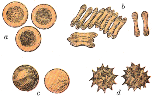
تتخذ خلايا الدم الحمراء لدى الثدييات عادةً شكل أقراص مقعرة الوجهين: مسطحة ومنخفضة في المركز، ذات مقطع عرضي يشبه الدمبل ، وحافة دائرية على طرف القرص. يسمح هذا الشكل بنسبة عالية بين مساحة السطح والحجم (SA/V) لتسهيل انتشار الغازات. [ 16 ] ومع ذلك، توجد بعض الاستثناءات فيما يتعلق بالشكل في رتبة مزدوجات الأصابع ( ذوات الحوافر زوجية الأصابع ، بما في ذلك الأبقار والغزلان وأقاربها)، والتي تُظهر مجموعة واسعة من أشكال خلايا الدم الحمراء الغريبة: خلايا صغيرة بيضاوية الشكل للغاية في اللاما والإبل ( فصيلة الجمليات )، وخلايا كروية صغيرة في غزلان الفأر (فصيلة الغزلان )، وخلايا تتخذ أشكالًا مغزلية ورمحية وهلالية ومضلعة غير منتظمة وأشكالًا زاوية أخرى في الأيل الأحمر والأيل الجبلي (فصيلة الأيائل ). من الواضح أن أفراد هذه الرتبة قد طوروا نمطًا لتكوين خلايا الدم الحمراء يختلف اختلافًا جوهريًا عن النمط الطبيعي لدى الثدييات. [ 10 ] [ 17 ] عمومًا، تتميز خلايا الدم الحمراء لدى الثدييات بمرونة وقابلية تشكيل ملحوظة تمكنها من المرور عبر الشعيرات الدموية الدقيقة ، فضلًا عن زيادة مساحة سطحها الملامس للسطح الخارجي من خلال اتخاذ شكل السيجار، حيث تطلق حمولتها من الأكسجين بكفاءة عالية. [ 18 ]
تتميز خلايا الدم الحمراء لدى الثدييات عن غيرها من الفقاريات، إذ لا تحتوي معظم الأنواع على نواة عند نضجها. [ 19 ] تحتوي هذه الخلايا على نواة خلال المراحل المبكرة من تكوينها ، لكنها تتخلص منها أثناء نموها ونضجها، مما يوفر مساحة أكبر للهيموجلوبين. وتفقد خلايا الدم الحمراء الخالية من النواة، والتي تُسمى الخلايا الشبكية ، جميع العضيات الخلوية الأخرى، مثل الميتوكوندريا وجهاز جولجي والشبكة الإندوبلازمية .
يعمل الطحال كمخزن لخلايا الدم الحمراء، لكن هذا التأثير محدود نوعًا ما لدى البشر. أما في بعض الثدييات الأخرى، كالكلاب والخيول، فيقوم الطحال باحتجاز أعداد كبيرة من خلايا الدم الحمراء، التي تُطلق في الدم أثناء فترات الإجهاد البدني، مما يُحسّن قدرة نقل الأكسجين .

بشر


يبلغ قطر قرص خلية الدم الحمراء البشرية النموذجية حوالي 6.2-8.2 ميكرومتر [ 20 ] ، ويبلغ أقصى سمك لها 2-2.5 ميكرومتر، بينما يبلغ أدنى سمك لها في المركز 0.8-1 ميكرومتر، وهي أصغر بكثير من معظم خلايا الدم البشرية الأخرى . يبلغ متوسط حجم هذه الخلايا حوالي 90 فمتولتر [ 21 ] ، ومساحة سطحها حوالي 136 ميكرومتر مربع ، ويمكنها أن تنتفخ لتصبح كروية الشكل تحتوي على 150 فمتولتر، دون تمدد الغشاء.
يحتوي جسم الإنسان البالغ على ما يقارب 20-30 تريليون خلية دم حمراء في أي وقت، ما يشكل حوالي 70% من إجمالي خلايا الدم. [ 22 ] ويبلغ عدد خلايا الدم الحمراء لدى النساء حوالي 4-5 ملايين خلية لكل ميكرولتر (ملليمتر مكعب) من الدم، بينما يبلغ لدى الرجال حوالي 5-6 ملايين خلية؛ أما الأشخاص الذين يعيشون في المرتفعات العالية ذات مستويات الأكسجين المنخفضة، فيكون لديهم عدد أكبر. وبالتالي، فإن خلايا الدم الحمراء أكثر شيوعًا من جزيئات الدم الأخرى: إذ يوجد حوالي 4000-11000 خلية دم بيضاء ، وحوالي 150000-400000 صفيحة دموية لكل ميكرولتر.
تستغرق خلايا الدم الحمراء البشرية في المتوسط 60 ثانية لإكمال دورة واحدة في الدورة الدموية. [ 5 ] [ 9 ] [ 23 ]
يعود اللون الأحمر للدم إلى الخصائص الطيفية لأيونات الحديد الهيمية في الهيموجلوبين . يحمل كل جزيء هيموجلوبين أربع مجموعات هيم، ويشكل الهيموجلوبين حوالي ثلث حجم الخلية الكلي. يُعد الهيموجلوبين مسؤولاً عن نقل أكثر من 98% من الأكسجين في الجسم (يُحمل الأكسجين المتبقي مذابًا في بلازما الدم ). تخزن خلايا الدم الحمراء لدى رجل بالغ متوسط الحجم حوالي 2.5 غرام من الحديد، وهو ما يمثل حوالي 65% من إجمالي الحديد الموجود في الجسم. [ 24 ] [ 25 ]
البنية المجهرية
نواة
Red blood cells in mammals are anucleate when mature, meaning that they lack a cell nucleus. In comparison, the red blood cells of other vertebrates have nuclei; the only known exceptions are salamanders of the family Plethodontidae, where five different clades has evolved various degrees of enucleated red blood cells (most evolved in some species of the genus Batrachoseps), and fish of the genus Maurolicus.[26][27][28]
The elimination of the nucleus in vertebrate red blood cells has been offered as an explanation for the subsequent accumulation of non-coding DNA in the genome.[17] The argument runs as follows: Efficient gas transport requires red blood cells to pass through very narrow capillaries, and this constrains their size. In the absence of nuclear elimination, the accumulation of repeat sequences is constrained by the volume occupied by the nucleus, which increases with genome size.
Nucleated red blood cells in mammals consist of two forms: normoblasts, which are normal erythropoietic precursors to mature red blood cells, and megaloblasts, which are abnormally large precursors that occur in megaloblastic anemias.
Membrane composition
Red blood cells are deformable, flexible, are able to adhere to other cells, and are able to interface with immune cells. Their membrane plays many roles in this. These functions are highly dependent on the membrane composition. The red blood cell membrane is composed of 3 layers: the glycocalyx on the exterior, which is rich in carbohydrates; the lipid bilayer which contains many transmembrane proteins, besides its lipidic main constituents; and the membrane skeleton, a structural network of proteins located on the inner surface of the lipid bilayer. Half of the membrane mass in human and most mammalian red blood cells are proteins. The other half are lipids, namely phospholipids and cholesterol.[29]
Membrane lipids

يتكون غشاء خلايا الدم الحمراء من طبقة ثنائية نموذجية من الدهون ، تشبه تلك الموجودة في جميع خلايا الجسم تقريبًا. ببساطة، تتكون هذه الطبقة الثنائية من الكوليسترول والفوسفوليبيدات بنسب متساوية وزنيًا. يُعدّ تركيب الدهون مهمًا لأنه يُحدد العديد من الخصائص الفيزيائية، مثل نفاذية الغشاء وسيولته. بالإضافة إلى ذلك، يتم تنظيم نشاط العديد من بروتينات الغشاء من خلال تفاعلاتها مع الدهون في الطبقة الثنائية.
على عكس الكوليسترول، الذي يتوزع بالتساوي بين الطبقتين الداخلية والخارجية، فإن الفوسفوليبيدات الخمسة الرئيسية موزعة بشكل غير متماثل، كما هو موضح أدناه:
الطبقة الخارجية الأحادية
- فوسفاتيديل كولين (PC)؛
- السفينغوميلين (SM).
الطبقة الداخلية الأحادية
- فوسفاتيديل إيثانولامين (PE)؛
- فوسفاتيديل إينوزيتول (PI) (كميات صغيرة).
- فوسفاتيديل سيرين (PS)؛
يُعزى هذا التوزيع غير المتماثل للفوسفوليبيدات بين طبقتي الغشاء إلى وظيفة العديد من بروتينات نقل الفوسفوليبيدات ، بعضها يعتمد على الطاقة والبعض الآخر لا يعتمد عليها. تنقل بروتينات تُسمى " فليباز " الفوسفوليبيدات من الطبقة الخارجية إلى الطبقة الداخلية، بينما تقوم بروتينات أخرى تُسمى " فلوباز " بالعملية المعاكسة، أي عكس تدرج التركيز بطريقة تعتمد على الطاقة. إضافةً إلى ذلك، توجد أيضًا بروتينات " سكرامبليز " التي تنقل الفوسفوليبيدات في كلا الاتجاهين في آنٍ واحد، وفقًا لتدرجات تركيزها بطريقة لا تعتمد على الطاقة. ولا يزال هناك جدل كبير قائم حول هوية هذه البروتينات المسؤولة عن صيانة غشاء خلايا الدم الحمراء.
يُعد الحفاظ على توزيع غير متماثل للفوسفوليبيدات في الطبقة الثنائية (مثل التموضع الحصري للفوسفاتيديل سيرين والفوسفاتيديل إينوزيتول في الطبقة الأحادية الداخلية) أمرًا بالغ الأهمية لسلامة الخلية ووظيفتها لعدة أسباب:
- تتعرف الخلايا البلعمية على خلايا الدم الحمراء التي تُظهر الفوسفاتيديل سيرين على سطحها الخارجي، وتقوم بابتلاعها . لذا، فإن حصر الفوسفاتيديل سيرين في الطبقة الأحادية الداخلية أمر ضروري لبقاء الخلية على قيد الحياة في مواجهة خلايا الجهاز الشبكي البطاني بشكل متكرر ، وخاصة في الطحال .
- وقد تم ربط التدمير المبكر لخلايا الدم الحمراء المصابة بالثلاسيميا والمنجلية باضطرابات عدم تناسق الدهون مما يؤدي إلى كشف PS على الطبقة الأحادية الخارجية.
- قد يؤدي تعرض الفوسفاتيديل سيرين (PS) إلى زيادة التصاق خلايا الدم الحمراء بالخلايا البطانية الوعائية، مما يعيق مرورها الطبيعي عبر الأوعية الدموية الدقيقة. لذا، من المهم الحفاظ على وجود الفوسفاتيديل سيرين في الطبقة الداخلية فقط من الغشاء ثنائي الطبقة لضمان تدفق الدم الطبيعي في الدورة الدموية الدقيقة.
- Both PS and phosphatidylinositol 4,5-bisphosphate (PIP2) can regulate membrane mechanical function, due to their interactions with skeletal proteins such as spectrin and protein 4.1R. Recent studies have shown that binding of spectrin to PS promotes membrane mechanical stability. PIP2 enhances the binding of protein band 4.1R to glycophorin C but decreases its interaction with protein band 3, and thereby may modulate the linkage of the bilayer to the membrane skeleton.
The presence of specialized structures named "lipid rafts" in the red blood cell membrane have been described by recent studies. These are structures enriched in cholesterol and sphingolipids associated with specific membrane proteins, namely flotillins, STOMatins (band 7), G-proteins, and β-adrenergic receptors. Lipid rafts that have been implicated in cell signaling events in nonerythroid cells have been shown in erythroid cells to mediate β2-adregenic receptor signaling and increase cAMP levels, and thus regulating entry of malarial parasites into normal red cells.[30][31]
Membrane proteins

The proteins of the membrane skeleton are responsible for the deformability, flexibility and durability of the red blood cell, enabling it to squeeze through capillaries less than half the diameter of the red blood cell (7–8 μm) and recovering the discoid shape as soon as these cells stop receiving compressive forces, in a similar fashion to an object made of rubber.
There are currently more than 50 known membrane proteins, which can exist in a few hundred up to a million copies per red blood cell. Approximately 25 of these membrane proteins carry the various blood group antigens, such as the A, B and Rh antigens, among many others. These membrane proteins can perform a wide diversity of functions, such as transporting ions and molecules across the red cell membrane, adhesion and interaction with other cells such as endothelial cells, as signaling receptors, as well as other currently unknown functions. The blood types of humans are due to variations in surface glycoproteins of red blood cells. Disorders of the proteins in these membranes are associated with many disorders, such as hereditary spherocytosis, hereditary elliptocytosis, hereditary stomatocytosis, and paroxysmal nocturnal hemoglobinuria.[29][30]
The red blood cell membrane proteins organized according to their function:

Transport
- Band 3 – Anion transporter, also an important structural component of the red blood cell membrane, makes up to 25% of the cell membrane surface, each red cell contains approximately one million copies. Defines the Diego Blood Group;[33]
- Aquaporin 1 – water transporter, defines the Colton Blood Group;
- Glut1 – glucose and L-dehydroascorbic acid transporter;
- MCT1 – Monocarboxylate transporter for exporting Lactic acid to the liver. See Cori cycle.;[34]
- Kidd antigen protein – urea transporter;
- RHAG – gas transporter, probably of carbon dioxide, defines Rh Blood Group and the associated unusual blood group phenotype Rhnull;
- Na+/K+ – ATPase;
- Ca2+ – ATPase;
- Na+ K+ 2Cl− – cotransporter;
- Na+-Cl− – cotransporter;
- Na-H exchanger;
- K-Cl – cotransporter;
- Gardos Channel.
Cell adhesion
- ICAM-4 – interacts with integrins;
- BCAM – a glycoprotein that defines the Lutheran blood group and also known as Lu or laminin-binding protein.
الدور الهيكلي - تعمل البروتينات الغشائية التالية على إنشاء روابط مع البروتينات الهيكلية وقد تلعب دورًا مهمًا في تنظيم التماسك بين الطبقة الدهنية الثنائية والهيكل الغشائي، مما قد يمكّن الخلية الحمراء من الحفاظ على مساحة سطح الغشاء المواتية عن طريق منع الغشاء من الانهيار (التكيس).
- مركب جزيئي ضخم قائم على الأنكيرين - بروتينات تربط الطبقة الثنائية بالهيكل الغشائي من خلال تفاعل نطاقاتها السيتوبلازمية مع الأنكيرين .
- يقوم النطاق 3 أيضًا بتجميع العديد من إنزيمات التحلل السكري ، وناقل ثاني أكسيد الكربون المفترض ، وأنهيدراز الكربونيك في مركب جزيئي ضخم يسمى " ميتابولون "، والذي قد يلعب دورًا رئيسيًا في تنظيم استقلاب خلايا الدم الحمراء ووظيفة نقل الأيونات والغازات .
- RHAG - يشارك أيضًا في النقل، ويحدد النمط الظاهري غير العادي المرتبط بفصيلة الدم Rh mod .
- مركب جزيئي ضخم قائم على البروتين 4.1R - البروتينات التي تتفاعل مع البروتين 4.1R .
- البروتين 4.1R – تعبير ضعيف عن مستضدات جرثومة الجرب ؛
- بروتين جليكوبروتين C و D – بروتين سكري، يحدد فصيلة دم جيربيتش ؛
- XK – يحدد فصيلة دم كيل والنمط الظاهري غير العادي لماكلويد (نقص مستضد Kx وانخفاض كبير في التعبير عن مستضدات كيل)؛
- RhD/RhCE – يحدد فصيلة الدم Rh والنمط الظاهري غير العادي المرتبط بها Rh null ؛
- تم اقتراح أن بروتين دافي مرتبط بتصفية الكيموكين ؛ [ 35 ]
- أدكتين – التفاعل مع النطاق 3؛
- ديماتين - التفاعل مع ناقل الجلوكوز Glut1.
الجهد الكهروستاتيكي السطحي
جهد زيتا هو خاصية كهروكيميائية لأسطح الخلايا، ويتحدد بالشحنة الكهربائية الصافية للجزيئات المعرضة على سطح أغشية الخلايا. يبلغ جهد زيتا الطبيعي لخلايا الدم الحمراء -15.7 ملي فولت (mV). [ 36 ] ويبدو أن جزءًا كبيرًا من هذا الجهد ناتج عن بقايا حمض السياليك المكشوفة في الغشاء: إذ يؤدي إزالتها إلى جهد زيتا قدره -6.06 ملي فولت.
وظيفة
دورها في نقل ثاني أكسيد الكربون
تذكر أن عملية التنفس ، كما هو موضح تخطيطيًا هنا بوحدة من الكربوهيدرات، تنتج عددًا من جزيئات ثاني أكسيد الكربون، CO2، يساوي تقريبًا عدد جزيئات الأكسجين، O2 . [ 37 ]
![{\displaystyle {\mathrm {HCOH} {}+{}\mathrm {O} {\vphantom {A}}_{\smash[{t}]{2}}{}\mathrel {\longrightarrow } {}\mathrm {CO} {\vphantom {A}}_{\smash[{t}]{2}}{}+{}\mathrm {H} {\vphantom {A}}_{\smash[{t}]{2}}\mathrm {O} }}](https://wikimedia.org/api/rest_v1/media/math/render/svg/b27b28b7948903c901385e2a8c71b5967168e8b9)
وبالتالي، فإن وظيفة الجهاز الدوري لا تقل أهمية عن نقل الأكسجين، فهي لا تقتصر على نقل ثاني أكسيد الكربون فحسب، بل تشمل نقل الأكسجين أيضاً. وكما ذُكر في موضع آخر من هذه المقالة، فإن معظم ثاني أكسيد الكربون في الدم يكون على شكل أيون البيكربونات، الذي يُوفر مُنظماً حرجاً لدرجة الحموضة . [ 38 ] لذا، وعلى عكس الهيموجلوبين المسؤول عن نقل الأكسجين ، فإن عدم وجود جزيء ناقل مُخصص لثاني أكسيد الكربون يُعد ميزة فسيولوجية .
مع ذلك، تلعب خلايا الدم الحمراء دورًا محوريًا في عملية نقل ثاني أكسيد الكربون ، وذلك لسببين. أولًا، لأنها، بالإضافة إلى الهيموجلوبين، تحتوي على عدد كبير من نسخ إنزيم الأنهيدراز الكربوني على الغشاء الخلوي الداخلي. [ 39 ] يعمل الأنهيدراز الكربوني، كما يوحي اسمه، كمحفز لتبادل حمض الكربونيك وثاني أكسيد الكربون (وهو أنهيدريد حمض الكربونيك). ولأنه محفز، فإنه يؤثر على العديد من جزيئات ثاني أكسيد الكربون ، وبالتالي يؤدي دوره الأساسي دون الحاجة إلى عدد كبير من النسخ كما هو الحال لنقل الأكسجين بواسطة الهيموجلوبين. في وجود هذا المحفز، يصل ثاني أكسيد الكربون وحمض الكربونيك إلى حالة توازن بسرعة كبيرة، بينما لا تزال خلايا الدم الحمراء تتحرك عبر الشعيرات الدموية. وهكذا، تضمن خلايا الدم الحمراء نقل معظم ثاني أكسيد الكربون على شكل بيكربونات . [ 40 ] [ 41 ] عند درجة الحموضة الفسيولوجية، يميل التوازن بشدة لصالح حمض الكربونيك، الذي يتفكك في الغالب إلى أيون البيكربونات. [ 42 ]
![{\displaystyle {\mathrm {CO} {\vphantom {A}}_{\smash[{t}]{2}}{}+{}\mathrm {H} {\vphantom {A}}_{\smash[{t}]{2}}\mathrm {O} {}\mathrel {\longRightleftharpoons } {}\mathrm {H} {\vphantom {A}}_{\smash[{t}]{2}}\mathrm {CO} {\vphantom {A}}_{\smash[{t}]{3}}{}\mathrel {\longRightleftharpoons } {}\mathrm {HCO} {\vphantom {A}}_{\smash[{t}]{3}}{\vphantom {A}}^{-}{}+{}\mathrm {H} {\vphantom {A}}^{+}}}](https://wikimedia.org/api/rest_v1/media/math/render/svg/d3c042753831a3fb66d69113b9daf9d889514778)
تعمل أيونات H+ التي يتم إطلاقها من خلال هذا التفاعل السريع داخل خلايا الدم الحمراء، بينما لا تزال في الشعيرات الدموية، على تقليل ألفة ارتباط الأكسجين بالهيموجلوبين، وهو تأثير بور .
تتمثل المساهمة الرئيسية الثانية لخلايا الدم الحمراء في نقل ثاني أكسيد الكربون في تفاعله المباشر مع مكونات بروتين الغلوبين في الهيموغلوبين لتكوين مركبات كاربامينوهيموغلوبين . ومع إطلاق الأكسجين في الأنسجة، يرتبط المزيد من ثاني أكسيد الكربون بالهيموغلوبين ، وعندما يرتبط الأكسجين في الرئتين، فإنه يحل محل ثاني أكسيد الكربون المرتبط بالهيموغلوبين ، وهذا ما يُعرف بتأثير هالدين . وعلى الرغم من أن كمية ضئيلة فقط من ثاني أكسيد الكربون في الدم ترتبط بالهيموغلوبين في الدم الوريدي، فإن نسبة أكبر من التغير في محتوى ثاني أكسيد الكربون بين الدم الوريدي والشرياني ناتجة عن التغير في هذا ثاني أكسيد الكربون المرتبط . [ 43 ] أي أن هناك وفرة دائمة من البيكربونات في الدم، سواء الوريدي أو الشرياني، نظرًا لدورها المذكور سابقًا كمنظم لدرجة الحموضة.
باختصار، ينتشر ثاني أكسيد الكربون الناتج عن التنفس الخلوي بسرعة كبيرة إلى مناطق ذات تركيز أقل، وتحديدًا إلى الشعيرات الدموية المجاورة. [ 44 ] [ 45 ] عند انتشاره إلى داخل خلية الدم الحمراء، يتحول ثاني أكسيد الكربون بسرعة بواسطة إنزيم الأنهيدراز الكربوني الموجود على الغشاء الداخلي للخلية إلى أيون البيكربونات. تغادر أيونات البيكربونات بدورها خلية الدم الحمراء مقابل أيونات الكلوريد من البلازما، وذلك بفضل بروتين نقل الأنيونات من النوع 3 الموجود في غشاء خلية الدم الحمراء. لا يعود أيون البيكربونات إلى خارج الشعيرة الدموية، بل يُنقل إلى الرئتين. في الرئتين، يؤدي انخفاض الضغط الجزئي لثاني أكسيد الكربون في الحويصلات الهوائية إلى انتشاره بسرعة من الشعيرة الدموية إلى الحويصلات الهوائية. يحافظ إنزيم الأنهيدراز الكربوني في خلايا الدم الحمراء على توازن أيون البيكربونات مع ثاني أكسيد الكربون. لذا، عندما يخرج ثاني أكسيد الكربون من الشعيرة الدموية، ويحل الأكسجين محل ثاني أكسيد الكربون على الهيموجلوبين، يتحول أيون البيكربونات بكمية كافية وبسرعة إلى ثاني أكسيد الكربون للحفاظ على التوازن. [ 39 ] [ 46 ] [ 47 ] [ 48 ]
الوظائف الثانوية
عندما تتعرض خلايا الدم الحمراء لإجهاد القص في الأوعية الدموية المتضيقة، فإنها تطلق الأدينوسين ثلاثي الفوسفات (ATP )، مما يؤدي إلى استرخاء جدران الأوعية الدموية وتوسعها لتعزيز تدفق الدم الطبيعي. [ 49 ]
عندما يتم إزالة الأكسجين من جزيئات الهيموجلوبين الخاصة بها، تطلق خلايا الدم الحمراء S-Nitrosothiols ، والتي تعمل أيضًا على توسيع الأوعية الدموية، [ 50 ] وبالتالي توجيه المزيد من الدم إلى مناطق الجسم التي تعاني من نقص الأكسجين.
تستطيع خلايا الدم الحمراء أيضاً تصنيع أكسيد النيتريك إنزيمياً، باستخدام الأرجينين كمادة أساسية، كما تفعل الخلايا البطانية . [ 51 ] يؤدي تعريض خلايا الدم الحمراء لمستويات فسيولوجية من إجهاد القص إلى تنشيط إنزيم أكسيد النيتريك سينثاز وتصدير أكسيد النيتريك، [ 52 ] مما قد يساهم في تنظيم توتر الأوعية الدموية.
تستطيع خلايا الدم الحمراء أيضاً إنتاج كبريتيد الهيدروجين ، وهو غاز إشاري يعمل على إرخاء جدران الأوعية الدموية. ويُعتقد أن التأثيرات الوقائية للثوم على القلب تعود إلى تحويل خلايا الدم الحمراء لمركبات الكبريت الموجودة فيه إلى كبريتيد الهيدروجين. [ 53 ]
تؤدي خلايا الدم الحمراء أيضاً دوراً في استجابة الجسم المناعية : فعندما تتحلل بفعل مسببات الأمراض مثل البكتيريا، يطلق الهيموجلوبين الموجود فيها جذوراً حرة ، والتي بدورها تُحلل جدار الخلية والغشاء الخاص بمسبب المرض، مما يؤدي إلى قتله. [ 54 ] [ 55 ]
العمليات الخلوية
نتيجةً لعدم احتوائها على الميتوكوندريا ، لا تستخدم خلايا الدم الحمراء أيًا من الأكسجين الذي تنقله؛ بل تُنتج جزيء ATP، وهو ناقل الطاقة ، من خلال تحلل الجلوكوز وتخمر حمض اللاكتيك على البيروفات الناتج . [ 56 ] [ 57 ] علاوة على ذلك، يلعب مسار فوسفات البنتوز دورًا هامًا في خلايا الدم الحمراء؛ انظر نقص إنزيم نازعة هيدروجين الجلوكوز-6-فوسفات لمزيد من المعلومات.
بما أن خلايا الدم الحمراء لا تحتوي على نواة، فمن المفترض حاليًا أن عملية تخليق البروتين غائبة في هذه الخلايا.
Because of the lack of nuclei and organelles, mature red blood cells do not contain DNA and cannot synthesize any RNA (although it does contain RNAs),[58][59] and consequently cannot divide and have limited repair capabilities.[60] The inability to carry out protein synthesis means that no virus can evolve to target mammalian red blood cells.[61] However, infection with parvoviruses (such as human parvovirus B19) can affect erythroid precursors while they still have DNA, as recognized by the presence of giant pronormoblasts with viral particles and inclusion bodies, thus temporarily depleting the blood of reticulocytes and causing anemia.[62]
Life cycle
Human red blood cells are produced through a process named erythropoiesis, developing from committed stem cells to mature red blood cells in about 7 days. When matured, in a healthy individual these cells live in blood circulation for about 100 to 120 days (and 80 to 90 days in a full term infant).[63] At the end of their lifespan, they are removed from circulation. In many chronic diseases, the lifespan of the red blood cells is reduced.
Creation
Erythropoiesis is the process by which new red blood cells are produced; it lasts about 7 days. Through this process red blood cells are continuously produced in the red bone marrow of large bones. (In the embryo, the liver is the main site of red blood cell production.) The production can be stimulated by the hormoneerythropoietin (EPO), synthesised by the kidney. Just before and after leaving the bone marrow, the developing cells are known as reticulocytes; these constitute about 1% of circulating red blood cells.
Functional lifetime
The functional lifetime of a red blood cell is about 100–120 days, during which time the red blood cells are continually moving in the circulatory system.[64]
Senescence
The aging red blood cell undergoes changes in its plasma membrane, making it susceptible to selective recognition by macrophages and subsequent phagocytosis in the mononuclear phagocyte system (spleen, liver and lymph nodes), thus removing old and defective cells and continually purging the blood. This process is termed eryptosis, red blood cell programmed death.[65] This process normally occurs at the same rate of production by erythropoiesis, balancing the total circulating red blood cell count. Eryptosis is increased in a wide variety of diseases including sepsis, haemolytic uremic syndrome, malaria, sickle cell anemia, beta-thalassemia, glucose-6-phosphate dehydrogenase deficiency, phosphate depletion, iron deficiency and Wilson's disease. Eryptosis can be elicited by osmotic shock, oxidative stress, and energy depletion, as well as by a wide variety of endogenous mediators and xenobiotics. Excessive eryptosis is observed in red blood cells lacking the cGMP-dependent protein kinase type I or the AMP-activated protein kinase AMPK. Inhibitors of eryptosis include erythropoietin, nitric oxide, catecholamines and high concentrations of urea.
Much of the resulting breakdown products are recirculated in the body. The heme constituent of hemoglobin are broken down into iron (Fe3+) and biliverdin. The biliverdin is reduced to bilirubin, which is released into the plasma and recirculated to the liver bound to albumin. The iron is released into the plasma to be recirculated by a carrier protein called transferrin. Almost all red blood cells are removed in this manner from the circulation before they are old enough to hemolyze. Hemolyzed hemoglobin is bound to a protein in plasma called haptoglobin, which is not excreted by the kidney.[66]
Clinical significance
Disease
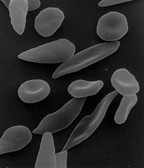
Blood diseases involving the red blood cells include:
- Anemias (or anaemias) are diseases characterized by low oxygen transport capacity of the blood, because of low red cell count or some abnormality of the red blood cells or the hemoglobin.
- Iron deficiency anemia is the most common anemia; it occurs when the dietary intake or absorption of iron is insufficient, and hemoglobin, which contains iron, cannot be formed.
- Pernicious anemia is an autoimmune disease wherein the body lacks intrinsic factor, required to absorb vitamin B12 from food. Vitamin B12 is needed for the production of red blood cells and hemoglobin.
- Sickle-cell disease is a genetic disease that results in abnormal hemoglobin molecules. When these release their oxygen load in the tissues, they become insoluble, leading to mis-shaped red blood cells. These sickle shaped red cells are less deformable and viscoelastic, meaning that they have become rigid and can cause blood vessel blockage, pain, strokes, and other tissue damage.
- Thalassemia is a genetic disease that results in the production of an abnormal ratio of hemoglobin subunits.
- Hereditary spherocytosis syndromes are a group of inherited disorders characterized by defects in the red blood cell's cell membrane, causing the cells to be small, sphere-shaped, and fragile instead of donut-shaped and flexible. These abnormal red blood cells are destroyed by the spleen. Several other hereditary disorders of the red blood cell membrane are known.[67]
- Aplastic anemia is caused by the inability of the bone marrow to produce blood cells.
- Pure red cell aplasia is caused by the inability of the bone marrow to produce only red blood cells.


- Hemolysis is the general term for excessive breakdown of red blood cells. It can have several causes and can result in hemolytic anemia.
- The malaria parasite spends part of its life-cycle in red blood cells, feeds on their hemoglobin and then breaks them apart, causing fever. Both sickle-cell disease and thalassemia are more common in malaria areas, because these mutations convey some protection against the parasite.
- Polycythemias (or erythrocytoses) are diseases characterized by a surplus of red blood cells. The increased viscosity of the blood can cause a number of symptoms.
- In polycythemia vera the increased number of red blood cells results from an abnormality in the bone marrow.
- Several microangiopathic diseases, including disseminated intravascular coagulation and thrombotic microangiopathies, present with pathognomonic (diagnostic) red blood cell fragments called schistocytes. These pathologies generate fibrin strands that sever red blood cells as they try to move past a thrombus.
Transfusion
Red blood cells may be given as part of a blood transfusion. Blood may be donated from another person, or stored by the recipient at an earlier date. Donated blood usually requires screening to ensure that donors do not contain risk factors for the presence of blood-borne diseases, or will not suffer themselves by giving blood. Blood is usually collected and tested for common or serious blood-borne diseases including Hepatitis B, Hepatitis C and HIV. The blood type (A, B, AB, or O) or the blood product is identified and matched with the recipient's blood to minimise the likelihood of acute hemolytic transfusion reaction, a type of transfusion reaction. This relates to the presence of antigens on the cell's surface. After this process, the blood is stored, and within a short duration is used. Blood can be given as a whole product or the red blood cells separated as packed red blood cells.
Blood is often transfused when there is known anaemia, active bleeding, or when there is an expectation of serious blood loss, such as prior to an operation. Before blood is given, a small sample of the recipient's blood is tested with the transfusion in a process known as cross-matching.
In 2008 it was reported that human embryonic stem cells had been successfully coaxed into becoming red blood cells in the lab. The difficult step was to induce the cells to eject their nucleus; this was achieved by growing the cells on stromal cells from the bone marrow. It is hoped that these artificial red blood cells can eventually be used for blood transfusions.[68]
A human trial is conducted in 2022, using blood cultured from stem cells obtained from donor blood.[69]
Tests

Several blood tests involve red blood cells. These include a RBC count (the number of red blood cells per volume of blood), calculation of the hematocrit (percentage of blood volume occupied by red blood cells), and the erythrocyte sedimentation rate. The blood type needs to be determined to prepare for a blood transfusion or an organ transplantation.
Many diseases involving red blood cells are diagnosed with a blood film (or peripheral blood smear), where a thin layer of blood is smeared on a microscope slide. This may reveal poikilocytosis, which are variations in red blood cell shape. When red blood cells sometimes occur as a stack, flat side next to flat side. This is known as rouleaux formation, and it occurs more often if the levels of certain serum proteins are elevated, as for instance during inflammation.
Separation and blood doping
Red blood cells can be obtained from whole blood by centrifugation, which separates the cells from the blood plasma in a process known as blood fractionation. Packed red blood cells, which are made in this way from whole blood with the plasma removed, are used in transfusion medicine.[70] During plasma donation, the red blood cells are pumped back into the body right away and only the plasma is collected.
Some athletes have tried to improve their performance by blood doping: first about 1 litre of their blood is extracted, then the red blood cells are isolated, frozen and stored, to be reinjected shortly before the competition. (Red blood cells can be conserved for 5 weeks at −79 °C or −110 °F, or over 10 years using cryoprotectants[71]) This practice is hard to detect but may endanger the human cardiovascular system which is not equipped to deal with blood of the resulting higher viscosity. Another method of blood doping involves injection with erythropoietin to stimulate production of red blood cells. Both practices are banned by the World Anti-Doping Agency.
History
The first person to describe red blood cells was the young Dutch biologist Jan Swammerdam, who had used an early microscope in 1658 to study the blood of a frog.[72] Unaware of this work, Anton van Leeuwenhoek provided another microscopic description in 1674, this time providing a more precise description of red blood cells, even approximating their size, "25,000 times smaller than a fine grain of sand".
In the 1740s, Vincenzo Menghini in Bologna was able to demonstrate the presence of iron by passing magnets over the powder or ash remaining from heated red blood cells.
In 1901, Karl Landsteiner published his discovery of the three main blood groups—A, B, and C (which he later renamed to O). Landsteiner described the regular patterns in which reactions occurred when serum was mixed with red blood cells, thus identifying compatible and conflicting combinations between these blood groups. A year later Alfred von Decastello and Adriano Sturli, two colleagues of Landsteiner, identified a fourth blood group—AB.
In 1959, by use of X-ray crystallography, Max Perutz was able to unravel the structure of hemoglobin, the red blood cell protein that carries oxygen.[73]
The oldest intact red blood cells ever discovered were found in Ötzi the Iceman, a natural mummy of a man who died around 3255 BCE. These cells were discovered in May 2012.[74]
See also
References
- ↑Kumar V, Abbas AK, Fausto N, Mitchell RN (2007). Robbins Basic Pathology (8th ed.). Saunders.
- ↑"Blood Cells". Archived from the original on 23 July 2016.
- ↑D'Alessandro A, Dzieciatkowska M, Nemkov T, Hansen KC (March 2017). "Red blood cell proteomics update: is there more to discover?". Blood Transfusion = Trasfusione del Sangue. 15 (2): 182–187. doi:10.2450/2017.0293-16. PMC 5336341. PMID 28263177.
- ↑Erich Sackmann, Biological Membranes Architecture and Function., Handbook of Biological Physics, (ed. R.Lipowsky and E.Sackmann, vol.1, Elsevier, 1995
- 1 2 بلوم، جيه إيه (2003). مراقبة التنفس والدورة الدموية . مطبعة سي آر سي. ص 27. ISBN 978-0-203-50328-7.
- ↑ هاتون، آي. إيه.، غالبريث، إي. دي.، ميرلو، إن. إس.، ميتينين، تي. بي.، سميث، بي. إم.، شاندر، جيه. إيه. (سبتمبر 2023). " عدد الخلايا البشرية وتوزيع أحجامها" . وقائع الأكاديمية الوطنية للعلوم في الولايات المتحدة الأمريكية . 120 (39) e2303077120. Bibcode : 2023PNAS..12003077H . doi : 10.1073/pnas.2303077120 . PMC 10523466. PMID 37722043 .
- ↑ سيندر ر، فوكس س، ميلو ر (أغسطس 2016). "تقديرات منقحة لعدد الخلايا البشرية والبكتيرية في الجسم" . مجلة PLOS Biology . 14 (8) e1002533. doi : 10.1371/journal.pbio.1002533 . PMC 4991899. PMID 27541692 .
- ↑ دين إل (2005). فصائل الدم ومستضدات خلايا الدم الحمراء . المركز الوطني لمعلومات التكنولوجيا الحيوية (الولايات المتحدة).
- 1 2 بييريج إف، سيرافيني إس، روسي إل، ماغناني إم (يناير 2008). "توصيل الأدوية باستخدام الخلايا". مراجعات توصيل الأدوية المتقدمة . 60 (2): 286-295 . doi : 10.1016/j.addr.2007.08.029 . PMID 17997501 .
- 1 2 غوليفر ج (1875). "حول حجم وشكل كريات الدم الحمراء في الفقاريات، مع رسومات توضيحية لها بمقياس موحد، وجداول قياسات موسعة ومنقحة". وقائع الجمعية الحيوانية في لندن . 1875 : 474-495 .
- ↑ رود ، ج. ت. (مايو 1954). "فقاريات بدون كريات دم حمراء وصبغة دموية". مجلة نيتشر . 173 (4410): 848-850 . Bibcode : 1954Natur.173..848R . doi : 10.1038/173848a0 . PMID 13165664. S2CID 3261779 .
- ↑ كارول س (2006). صناعة الأصلح . دبليو دبليو نورتون. رقم ISBN 978-0-393-06163-5.
- ↑ ماتون أ، هوبكنز ج، ماكلولين سي دبليو، جونسون إس، وارنر إم كيو، لاهارت دي، رايت جيه دي (1993). علم الأحياء البشري والصحة . إنجلوود كليفس، نيو جيرسي: برنتيس هول. ISBN 978-0-13-981176-0.
- ↑ أنثيس ن (17 أبريل 2008). "لماذا الأوردة زرقاء؟" . مدونات العلوم . تم الاطلاع عليه بتاريخ 23 أبريل 2015 .
- ↑ سنايدر جي كي، وشيفور بي إيه (1999). "خلايا الدم الحمراء: محور تطور الجهاز الدوري للفقاريات" . علم الأحياء التكاملي والمقارن . 39 (2): 189-198 . doi : 10.1093/icb/39.2.189 .
- ↑ "BBC Bitesize – GCSE Biology – Blood – Revision 2" . www.bbc.co.uk. تاريخ الاطلاع: 26 نوفمبر 2017 .
- 1 2 غريغوري تي آر (2001). "كلما زادت قيمة C، زاد حجم الخلية: حجم الجينوم وحجم خلايا الدم الحمراء في الفقاريات". خلايا الدم، الجزيئات والأمراض . 27 (5): 830-843 . CiteSeerX 10.1.1.22.9555 . doi : 10.1006/bcmd.2001.0457 . PMID 11783946 .
- ↑ غودمان إس آر، كورديا إيه، أمان إل، كاخنياشفيلي دي، دايسكو أو (ديسمبر 2007). "بروتينات وتفاعلات خلايا الدم الحمراء البشرية". علم الأحياء التجريبي والطب . 232 (11): 1391-1408 . doi : 10.3181/0706-MR-156 . PMID 18040063. S2CID 32326166 .
- ↑ ليبش، هانز-جورج، محرر (2019). علم الأنسجة البيطري للثدييات والطيور الأليفة . أطالس بيطرية ( الطبعة الخامسة). 5m Books Ltd. ISBN 9781789181067.
- ↑ تورجيون، إم إل (2004). علم الدم السريري: النظرية والإجراءات . ليبينكوت ويليامز وويلكنز. ص 100. ISBN 978-0-7817-5007-3.
- ↑ ماكلارين، سي إي، وبريتنهام، جي إم، وهاسيلبلاد، في (أبريل 1987). "التقييم الإحصائي والبياني لتوزيعات حجم كريات الدم الحمراء". المجلة الأمريكية لعلم وظائف الأعضاء . 252 (4 الجزء 2): H857– H866. CiteSeerX 10.1.1.1000.348 . doi : 10.1152/ajpheart.1987.252.4.H857 . PMID 3565597 .
- ↑Bianconi E, Piovesan A, Facchin F, Beraudi A, Casadei R, Frabetti F, et al. (1 November 2013). "An estimation of the number of cells in the human body". Annals of Human Biology. 40 (6): 463–471. doi:10.3109/03014460.2013.807878. hdl:11585/152451. PMID 23829164. S2CID 16247166.
- ↑Hillman RS, Ault KA, Rinder HM (2005). Hematology in Clinical Practice: A Guide to Diagnosis and Management (4th ed.). McGraw-Hill Professional. p. 1. ISBN 978-0-07-144035-6.
- ↑Iron Metabolism, University of Virginia Pathology. Accessed 22 September 2007.
- ↑Bridges KR. "Transferrin and Iron Transport Physiology". Information Center for Sickle Cell and Thalassemic Disorders. Retrieved 26 March 2023.
- ↑Cohen WD (1982). "The cytomorphic system of anucleate non-mammalian erythrocytes". Protoplasma. 113 (1): 23–32. Bibcode:1982Prpls.113...23C. doi:10.1007/BF01283036. S2CID 41287948.
- ↑Wingstrand KG (1956). "Non-nucleated erythrocytes in a teleostean fish Maurolicus mülleri (Gmelin)". Zeitschrift für Zellforschung und Mikroskopische Anatomie. 45 (2): 195–200. doi:10.1007/BF00338830. PMID 13402080. S2CID 12916049.
- ↑Mueller, R. L.; Gregory, T. R.; Gregory, S. M.; Hsieh, A.; Boore, J. L. (2008). "Genome size, cell size, and the evolution of enucleated erythrocytes in attenuate salamanders". Zoology. 111 (3): 218–230. Bibcode:2008Zool..111..218M. doi:10.1016/j.zool.2007.07.010. PMC 2435017. PMID 18328681.
- 1 2 3 يزدانبخش ك، لوماس-فرانسيس س، ريد إم إي (أكتوبر 2000). "فصائل الدم والأمراض المرتبطة بالتشوهات الوراثية في غشاء خلايا الدم الحمراء". مراجعات طب نقل الدم . 14 (4): 364-374 . doi : 10.1053/tmrv.2000.16232 . PMID 11055079 .
- 1 2 3 موهانداس ن، غالاغر ب.ج. (نوفمبر 2008). "غشاء خلايا الدم الحمراء: الماضي والحاضر والمستقبل" . مجلة الدم . 112 (10): 3939-3948 . doi : 10.1182 / blood-2008-07-161166 . PMC 2582001. PMID 18988878 .
- ↑ رودي، ب.م.، تروكو، ف.م.، جينارو، أ.م. (يونيو 2008). "العوامل المحددة لمقاومة أغشية كريات الدم الحمراء للمنظفات". الكيمياء الفيزيائية الحيوية . 135 ( 1-3 ): 14-18 . doi : 10.1016/j.bpc.2008.02.015 . hdl : 11336/24825 . PMID 18394774 .
- ↑ هيمبلمان إي، غوتزه أو (1984). "توصيف البروتينات الغشائية بواسطة التلوين الفضي متعدد الألوان". مجلة هوب-سيلر للكيمياء الفيزيولوجية . 365 : 241-242 .
- ↑ إيولاسكون أ، بيروتا س، ستيوارت ج.و. (مارس 2003). "عيوب غشاء خلايا الدم الحمراء". مراجعات في أمراض الدم السريرية والتجريبية . 7 (1): 22-56 . PMID 14692233 .
- ↑ فيسيل ب، شيفلر إي، شواب م (يوليو 2018). "الأهمية السريرية والوظيفية لعائلة ناقلات أحادي الكربوكسيلات في الفيزيولوجيا المرضية للأمراض والعلاج الدوائي" . العلوم السريرية والترجمية . 11 (4): 352-364 . doi : 10.1111/cts.12551 . PMC 6039204. PMID 29660777 .
- ↑ دينوم، جي إيه (يوليو 2004). "بنية ووظيفة الجزيئات التي تحمل مستضدات خلايا الدم الحمراء والصفائح الدموية البشرية". مراجعات طب نقل الدم . 18 (3): 203-231 . doi : 10.1016/j.tmrv.2004.03.006 . PMID 15248170 .
- ↑ توكوماسو ف، أوستيرا جي آر، أماراتونغا سي، فيرهيرست آر إم (2012) تعديلات في جهد زيتا لغشاء كريات الدم الحمراء نتيجة الإصابة بطفيل الملاريا المتصورة المنجلية . علم الطفيليات التجريبي
- ↑ جايتون، أ. س. (1976). "الفصل 41: نقل الأكسجين وثاني أكسيد الكربون في الدم وسوائل الجسم". كتاب علم وظائف الأعضاء الطبية ( الطبعة الخامسة). فيلادلفيا، بنسلفانيا: دبليو بي سوندرز. ص 556. ISBN 0-7216-4393-0
نسبة التبادل التنفسي هي 1:1 عند تناول الكربوهيدرات، وتكون منخفضة تصل إلى 0.7 عند تناول الدهون
. - ↑ ويست، ج. ب. (1974). "نقل الغازات إلى الأطراف". فسيولوجيا الجهاز التنفسي - الأساسيات . بالتيمور، ماريلاند: ويليامز وويلكنز. ص 80. ISBN 0-683-08932-3حالة التوازن الحمضي القاعدي :
يؤثر نقل ثاني أكسيد الكربون بشكل كبير على حالة التوازن الحمضي القاعدي في الدم والجسم ككل. تفرز الرئتان أكثر من 10000 ملي مكافئ من حمض الكربونيك يوميًا، مقارنةً بأقل من 100 ملي مكافئ من الأحماض الثابتة التي تفرزها الكلى.
- 1 2 جايتون، أ. س. (1976). "الفصل 41: نقل الأكسجين وثاني أكسيد الكربون في الدم وسوائل الجسم". كتاب علم وظائف الأعضاء الطبية ( الطبعة الخامسة). فيلادلفيا، بنسلفانيا: دبليو بي سوندرز. الصفحات 553-554 . ISBN 0-7216-4393-0
تفاعل ثاني أكسيد الكربون مع الماء في خلايا الدم الحمراء - تأثير الأنهيدراز
الكربوني - ↑ جايتون، أ. س. (1976). "الفصل 41: نقل الأكسجين وثاني أكسيد الكربون في الدم وسوائل الجسم". كتاب علم وظائف الأعضاء الطبية ( الطبعة الخامسة). فيلادلفيا، بنسلفانيا: دبليو بي سوندرز. الصفحات 553-554 . ISBN 0-7216-4393-0
يحفز إنزيم الأنهيدراز الكربوني التفاعل بين ثاني أكسيد الكربون والماء
. - ↑ كومرو الابن، جيه إتش (1965). "نقل وإخراج ثاني أكسيد الكربون". فسيولوجيا التنفس ( طبعة 1971). شيكاغو، إلينوي: دار نشر الكتاب الطبي السنوي. ص 176. ISBN 0-8151-1824-4إنزيم
[كربونيك أنهيدراز] يجعل التفاعل يسير نحو اليمين أسرع بحوالي 13000 مرة
- ↑ ديم ك، لينتنر سي، محرران (1970). "غازات الدم". جداول دوكومنتا جايجي العلمية ( الطبعة السابعة). بازل، سويسرا: سيبا-جايجي المحدودة. الصفحات 570-571 .
في البلازما، يوجد حوالي 5% من ثاني أكسيد الكربون في محلول فيزيائي، 94% منه على شكل بيكربونات و1% على شكل مركبات كاربامينية؛ أما في كريات الدم الحمراء، فالنسب المقابلة هي 7% و82% و11% على التوالي.
- ↑ جايتون، أ. س. (1976). "الفصل 41: نقل الأكسجين وثاني أكسيد الكربون في الدم وسوائل الجسم". كتاب علم وظائف الأعضاء الطبية ( الطبعة الخامسة). فيلادلفيا، بنسلفانيا: دبليو بي سوندرز. ص 554. ISBN 0-7216-4393-0.
from figure 41-5 Hgb.CO2 is about 23% and bicarbonate is about 70% of the total carbon dioxide transported to the lungs.
- ↑Comroe J (1965). "Pulmonary Gas Diffusion". Physiology of Respiration (1971 ed.). Chicago, IL: Year Book Medical Publishers. p. 140. ISBN 0-8151-1824-4.
Despite being a heavier molecule, because it is more soluble, the relative rate of diffusion of CO2 is about 20 times the rate of O2
- ↑Guyton AC (1976). "Ch. 41 Transport of Oxygen and Carbon Dioxide in the Blood and Body Fluids". Textbook of Medical Physiology (Fifth ed.). Philadelphia, PA: W. B. Saunders. p. 553. ISBN 0-7216-4393-0.
carbon dioxide diffuses out of the tissue cells in the gaseous form (but not to a significant effect in the bicarbonate form because the cell membrane is far less permeable to bicarbonate than to the dissolved gas.
- ↑Comroe Jr JH (1965). "Transport and elimination of carbon dioxide". Physiology of Respiration (1971 ed.). Chicago, IL: Year Book Medical Publishers. pp. 175–177. ISBN 0-8151-1824-4.
the buffering occurred in the red cell
- ↑West JB (1974). "Gas Transport to the Periphery". Respiratory Physiology – the essentials. Baltimore, MD: Williams & Wilkens. pp. 77–79. ISBN 0-683-08932-3.
CO2 Transport
- ↑Stone WE (1973). "Ch. 6-1 Uptake and Delivery of the Respiratory Gasses". In Brobeck JR (ed.). Best & Taylor's Physiological basis of medical practice (9th ed.). Baltimore, MD: Williams & Wilkins. pp. 6.16 –6.18. ISBN 0-683-10160-9.
Transport of CO2 as Bicarbonate
- ↑Wan J, Ristenpart WD, Stone HA (October 2008). "Dynamics of shear-induced ATP release from red blood cells". Proceedings of the National Academy of Sciences of the United States of America. 105 (43): 16432–16437. Bibcode:2008PNAS..10516432W. doi:10.1073/pnas.0805779105. PMC 2575437. PMID 18922780.
- ↑ ديزن دي إل، هيس دي تي، ستاملر جيه إس (أغسطس 2008). "توسع الأوعية الدموية الناتج عن نقص الأكسجين بواسطة خلايا الدم الحمراء: دليل على وجود إشارة تعتمد على إس-نيتروزوثيول" . أبحاث الدورة الدموية . 103 (5): 545-553 . doi : 10.1161/CIRCRESAHA.108.176867 . PMC 2763414. PMID 18658051 .
- ↑ كلاينبونغارد ب، شولز ر، راساف ت، لاور ت، ديجام أ، جاكس ت، وآخرون . (أبريل 2006). " خلايا الدم الحمراء تُعبّر عن إنزيم أكسيد النيتريك البطاني الوظيفي" . مجلة الدم . 107 (7): 2943-2951 . doi : 10.1182/blood-2005-10-3992 . PMID 16368881. S2CID 38270024 .
- ↑ أولكر، ب.، ساتي، ل.، سيليك-أوزينسي، ج.، ميسلمان، هـ. ج.، باسكرت، أ. ك. (2009). "التحفيز الميكانيكي لآليات تخليق أكسيد النيتريك في كريات الدم الحمراء". علم الريولوجيا الحيوية . 46 (2): 121-132 . doi : 10.3233/BIR-2009-0532 . PMID 19458415 .
- ↑ بينافيدس، جي. إيه.، سكوادريتو، جي. إل .، ميلز، آر. دبليو.، باتيل، إتش. دي.، إيزبيل، تي. إس.، باتيل، آر. بي.، وآخرون . (نوفمبر 2007). "كبريتيد الهيدروجين يُساهم في التأثير الوعائي للثوم" . وقائع الأكاديمية الوطنية للعلوم في الولايات المتحدة الأمريكية . 104 (46): 17977-17982 . Bibcode : 2007PNAS..10417977B . doi : 10.1073/pnas.0705710104 . PMC 2084282. PMID 17951430 .
- ↑ كيسافا س (1 سبتمبر 2007). "خلايا الدم الحمراء لا تقتصر وظيفتها على نقل الأكسجين فحسب؛ بل تُظهر نتائج جديدة لفريق من جامعة سنغافورة الوطنية أنها تهاجم البكتيريا بشراسة أيضًا" (ملف PDF) . صحيفة ستريتس تايمز . تاريخ الاطلاع: 26 مارس 2013 .
- ↑ جيانغ ن، تان ن س، هو ب، دينغ ج ل (أكتوبر 2007). "أنواع الأكسجين التفاعلية الناتجة عن بروتينات التنفس كاستراتيجية مضادة للميكروبات" . علم المناعة الطبيعي . 8 (10): 1114-1122 . doi : 10.1038/ni1501 . PMID 17721536. S2CID 11359246 .
- ^ بيرج جي إم، تيموكزكو جي إل، سترير إل (2012). الكيمياء الحيوية ( الطبعة السابعة). نيويورك: دبليو إتش فريمان. ص 455، 609. ردمك 978-1-4292-2936-4.
- ↑Tilton WM, Seaman C, Carriero D, Piomelli S (August 1991). "Regulation of glycolysis in the erythrocyte: role of the lactate/pyruvate and NAD/NADH ratios". The Journal of Laboratory and Clinical Medicine. 118 (2): 146–152. PMID 1856577.
- ↑Kabanova S, Kleinbongard P, Volkmer J, Andrée B, Kelm M, Jax TW (2009). "Gene expression analysis of human red blood cells". International Journal of Medical Sciences. 6 (4): 156–159. doi:10.7150/ijms.6.156. PMC 2677714. PMID 19421340.
- ↑Jain V, Yang WH, Wu J, Roback JD, Gregory SG, Chi JT (2022). "Single Cell RNA-Seq Analysis of Human Red Cells". Frontiers in Physiology. 13 828700. doi:10.3389/fphys.2022.828700. PMC 9065680. PMID 35514346.
- ↑Alberts, Bruce; Johnson, Alexander; Lewis, Julian; Raff, Martin; Roberts, Keith; Walter, Peter (2002). "Erythropoiesis Depends on the Hormone Erythropoietin". Molecular biology of the cell (4th ed.). New York: Garland. ISBN 0-8153-4072-9. Retrieved 30 November 2023.
- ↑Zimmer C (27 March 2007). "Scientists Explore Ways to Lure Viruses to Their Death". The New York Times. Retrieved 26 March 2013.
- ↑Heegaard ED, Brown KE (July 2002). "Human parvovirus B19". Clinical Microbiology Reviews. 15 (3): 485–505. doi:10.1128/CMR.15.3.485-505.2002. PMC 118081. PMID 12097253.
- ↑Harrison KL (June 1979). "Fetal erythrocyte lifespan". Australian Paediatric Journal. 15 (2): 96–97. doi:10.1111/j.1440-1754.1979.tb01197.x. PMID 485998. S2CID 5370064.
- ↑Higgins JM (March 2015). "Red blood cell population dynamics". Clinics in Laboratory Medicine. 35 (1): 43–57. doi:10.1016/j.cll.2014.10.002. PMC 4717490. PMID 25676371.
- ↑Lang F, Lang E, Föller M (October 2012). "Physiology and pathophysiology of eryptosis". Transfusion Medicine and Hemotherapy. 39 (5): 308–314. doi:10.1159/000342534. PMC 3678267. PMID 23801921.
- ↑Föller M, Huber SM, Lang F (October 2008). "Erythrocyte programmed cell death". IUBMB Life. 60 (10): 661–668. doi:10.1002/iub.106. PMID 18720418. S2CID 41603762.
- ↑An X, Mohandas N (May 2008). "Disorders of red cell membrane". British Journal of Haematology. 141 (3): 367–375. doi:10.1111/j.1365-2141.2008.07091.x. PMID 18341630. S2CID 7313716.
- ↑Coghlan A (19 August 2008). "First red blood cells grown in the lab". New Scientist. Retrieved 26 March 2023.
- ↑"Researchers are trialing lab-grown blood transfusions: What to know". medicalnewstoday.com. MedicalNewsToday. 11 November 2022. Archived from the original on 15 November 2022. Retrieved 17 November 2022.
A team of researchers led by the National Health Service (NHS) Blood and Transplant unit recently launched the first clinical trial to transfuse lab-grown red blood cells into a live human.
- ↑"Circular of Information for Blood and Blood Products"(PDF). American Association of Blood Banks, American Red Cross, America's Blood Centers. Archived from the original(PDF) on 30 October 2011. Retrieved 1 November 2010.
- ↑Valeri CR (8 February 1996). "Frozen Red Cell Technology". In Sparacino L, Manning FJ (eds.). Blood Groups and Red Cell Antigens. National Academies Press (US) – via www.ncbi.nlm.nih.gov.
- ↑"Swammerdam, Jan (1637–1680)", McGraw Hill AccessScience, 2007. Accessed 27 December 2007.
- ↑"Max F. Perutz – Biographical". NobelPrize.org. Retrieved 23 October 2018.
- ↑Pappas S (2 May 2012). "'Iceman' mummy holds world's oldest blood cells". Fox News. Archived from the original on 2 May 2012. Retrieved 2 May 2012.
External links
- Blood Groups and Red Cell Antigens by Laura Dean. Searchable and downloadable online textbook in the public domain.
- Database of vertebrate erythrocyte sizesArchived 5 July 2008 at the Wayback Machine.
- Red Gold, PBS site containing facts and history
- Human cells
- Blood cells
- Respiration
- 1658 in science