تُشير التقديرات إلى أن الكائنات الدقيقة البحرية تُشكّل ما بين 70 و90 بالمئة من الكتلة الحيوية في المحيط . وتُشكّل هذه الكائنات مجتمعةً ما يُعرف بالميكروبيوم البحري . وعلى مدى مليارات السنين، طوّر هذا الميكروبيوم أنماط حياة وتكيفات عديدة، وأصبح يُشارك في الدورة العالمية لجميع العناصر الكيميائية تقريبًا. [ 4 ] تُعدّ الكائنات الدقيقة بالغة الأهمية لإعادة تدوير المغذيات في النظم البيئية ، إذ تعمل كمحللات . كما أنها مسؤولة عن جميع عمليات التمثيل الضوئي تقريبًا التي تحدث في المحيط، بالإضافة إلى دورة الكربون والنيتروجين والفوسفور وغيرها من المغذيات والعناصر النزرة. [ 5 ] تُخزّن الكائنات الدقيقة البحرية كميات كبيرة من الكربون وتُنتج جزءًا كبيرًا من أكسجين العالم.
نسبة ضئيلة من الكائنات الدقيقة البحرية ممرضة ، وتسبب الأمراض، بل وحتى الموت، للنباتات والحيوانات البحرية. [ 6 ] مع ذلك، تعيد هذه الكائنات تدوير العناصر الكيميائية الرئيسية ، إذ تنتج وتستهلك نحو نصف إجمالي المواد العضوية المتولدة على كوكب الأرض سنويًا. وباعتبارها جزءًا من أكبر بيئة على وجه الأرض، تُحدث الأنظمة الميكروبية البحرية تغييرات في جميع الأنظمة العالمية.
في يوليو/تموز 2016، أعلن العلماء عن تحديد مجموعة من 355 جينًا من السلف المشترك الأخير (LUCA) لجميع أشكال الحياة على كوكب الأرض، بما في ذلك الكائنات الدقيقة البحرية. [ 7 ] وعلى الرغم من تنوعها، لا تزال الحياة المجهرية في المحيطات غير مفهومة بشكل كافٍ. فعلى سبيل المثال، لم يُستكشف دور الفيروسات في النظم البيئية البحرية إلا نادرًا حتى في بداية القرن الحادي والعشرين. [ 8 ]
تُعدّ الكائنات الدقيقة ضرورية لإعادة تدوير المغذيات في النظم البيئية ، إذ تعمل كمحللات . بعض هذه الكائنات ممرضة ، فتُسبب الأمراض، بل وحتى الموت، للنباتات والحيوانات. [ 6 ] وباعتبارها من سكان أكبر بيئة على وجه الأرض، تُحرك النظم البحرية الميكروبية التغيرات في كل نظام عالمي. تُعدّ الميكروبات مسؤولة عن جميع عمليات التمثيل الضوئي التي تحدث في المحيط تقريبًا، فضلًا عن دورة الكربون والنيتروجين والفوسفور وغيرها من المغذيات والعناصر النزرة. [ 5 ]
رغم التطورات التكنولوجية والاكتشافات العلمية الكبيرة التي شهدناها مؤخرًا، ما زلنا نفتقر إلى فهمٍ شاملٍ على جميع المستويات للأسئلة البيئية الأساسية المتعلقة بالكائنات الدقيقة في بحارنا ومحيطاتنا. وتتلخص هذه الأسئلة الجوهرية فيما يلي: 1. ما هي الكائنات الدقيقة الموجودة في بحارنا ومحيطاتنا، وما هو عددها؟ 2. ما هي وظائفها؟ ما هي الأدوار التي تؤديها كل من هذه الكائنات الدقيقة في البيئة البحرية، وكيف تُسهم في دورات الطاقة والمادة العالمية؟ 3. ما هي العوامل التي تُحدد وجود الكائنات الدقيقة أو غيابها، وكيف تؤثر على التنوع البيولوجي ووظائف الكائنات الدقيقة، والعكس صحيح؟
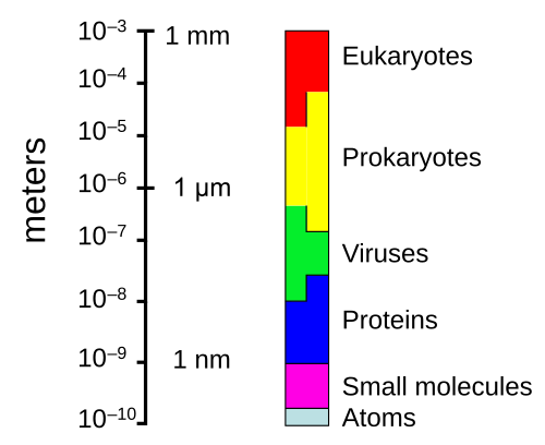
نطاق الأحجام التي تظهرها بدائيات النوى (البكتيريا والعتائق) والفيروسات مقارنة بأحجام الكائنات الحية الأخرى والجزيئات الحيوية.تحت عدسة مكبرة، تعج رذاذ مياه البحر بالحياة المجهرية
الحياة المجهرية تحت سطح البحر متنوعة، ولا يزال فهمها محدودًا، لا سيما فيما يتعلق بدور الفيروسات في النظم البيئية البحرية. [ 13 ] معظم الفيروسات البحرية هي عاثيات بكتيرية ، وهي غير ضارة بالنباتات والحيوانات، لكنها ضرورية لتنظيم النظم البيئية للمياه المالحة والعذبة. [ 14 ] تُصيب هذه العاثيات البكتيريا في المجتمعات الميكروبية المائية وتُدمرها، وهي الآلية الأهم لإعادة تدوير الكربون في البيئة البحرية. تُحفز الجزيئات العضوية المنبعثة من الخلايا البكتيرية الميتة نمو البكتيريا والطحالب الجديدة. [ 15 ] قد يُساهم النشاط الفيروسي أيضًا في المضخة البيولوجية ، وهي العملية التي يتم من خلالها عزل الكربون في أعماق المحيط. [ 16 ]
يمكن أن ينجرف رذاذ البحر الذي يحتوي على الكائنات الحية الدقيقة البحرية إلى طبقات الجو العليا حيث يصبح عوالق هوائية ، ويمكن أن يسافر حول العالم قبل أن يسقط عائدًا إلى الأرض.
تدور تيارات من الكائنات الدقيقة المحمولة جواً حول الكوكب فوق الأنظمة الجوية ولكن أسفل الممرات الجوية التجارية. [ 17 ] بعض هذه الكائنات الدقيقة المتنقلة تُحمل من عواصف الغبار الأرضية، لكن معظمها ينشأ من الكائنات الدقيقة البحرية في رذاذ البحر . في عام 2018، أفاد العلماء أن مئات الملايين من الفيروسات وعشرات الملايين من البكتيريا تترسب يومياً على كل متر مربع حول الكوكب. [ 18 ] [ 19 ]
تعيش الكائنات الحية الدقيقة في جميع أنحاء الغلاف الحيوي . قد تصل كتلة الكائنات الحية الدقيقة بدائية النواة - والتي تشمل البكتيريا والعتائق، ولكن ليس الكائنات الحية الدقيقة حقيقية النواة - إلى 0.8 تريليون طن من الكربون (من إجمالي كتلة الغلاف الحيوي ، المقدرة بما بين 1 و4 تريليونات طن). [ 20 ] وقد عُثر على ميكروبات بحرية وحيدة الخلية محبة للضغط العالي على عمق 10900 متر (35800 قدم) في خندق ماريانا ، أعمق نقطة في محيطات الأرض. [ 21 ] [ 22 ] تعيش الكائنات الدقيقة داخل الصخور على عمق 580 مترًا (1900 قدم) تحت قاع البحر، وعلى عمق 2590 مترًا (8500 قدم) من المحيط قبالة سواحل شمال غرب الولايات المتحدة ، [ 21 ] [ 23 ] وكذلك على عمق 2400 متر (7900 قدم؛ 1.5 ميل) تحت قاع البحر قبالة سواحل اليابان. [ 24 ] أعلى درجة حرارة معروفة يمكن أن تعيش فيها الكائنات الدقيقة هي 122 درجة مئوية (252 درجة فهرنهايت) ( Methanopyrus kandleri ). [ 25 ] في عام 2014، أكد العلماء وجود كائنات دقيقة تعيش على عمق 800 متر (2600 قدم) تحت جليد القارة القطبية الجنوبية . [ 26 ] [ 27 ] ووفقًا لأحد الباحثين، "يمكنك العثور على الميكروبات في كل مكان - فهي شديدة التكيف مع الظروف، وتعيش أينما وُجدت." [ 21 ] تُشكل الكائنات الدقيقة البحرية "أساس جميع السلاسل الغذائية البحرية، حيث تُعيد تدوير العناصر الرئيسية وتُنتج وتستهلك حوالي نصف المواد العضوية المتولدة على الأرض كل عام". [ 28 ] [ 29 ]
الفيروسات البحرية
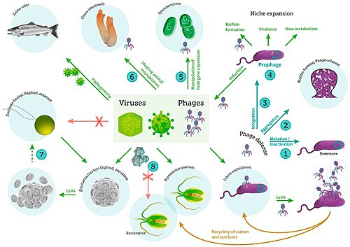
التفاعلات بين الفيروس والمضيف في النظام البيئي البحري، بما في ذلك العدوى الفيروسية للبكتيريا والعوالق النباتية والأسماك [ 30 ]
عندما لا تكون الفيروسات داخل خلية مصابة أو في طور إصابة خلية أخرى، فإنها توجد على شكل جسيمات مستقلة. تتكون هذه الجسيمات الفيروسية، المعروفة أيضًا باسم الفيروسات الكاملة ، من جزأين أو ثلاثة: (أ) المادة الوراثية (الجينوم) المصنوعة إما من الحمض النووي الريبوزي منقوص الأكسجين (DNA ) أو الحمض النووي الريبوزي ( RNA )، وهما جزيئان طويلان يحملان المعلومات الوراثية؛ (ب) غلاف بروتيني يُسمى القفيصة ، يُحيط بالمادة الوراثية ويحميها؛ وفي بعض الحالات (ج) غلاف من الدهون يُحيط بالغلاف البروتيني عندما تكون الفيروسات خارج الخلية. تتراوح أشكال هذه الجسيمات الفيروسية من الأشكال الحلزونية البسيطة والأشكال العشرين سطحية لبعض أنواع الفيروسات إلى تراكيب أكثر تعقيدًا لأنواع أخرى. تمتلك معظم أنواع الفيروسات فيروسات كاملة صغيرة جدًا بحيث لا يمكن رؤيتها بالمجهر الضوئي . يبلغ متوسط حجم الفيروس الكامل حوالي جزء من مئة من حجم البكتيريا المتوسطة .
لا تزال أصول الفيروسات في تاريخ تطور الحياة غير واضحة: فبعضها ربما تطور من البلازميدات - وهي أجزاء من الحمض النووي قادرة على الانتقال بين الخلايا - بينما تطور البعض الآخر من البكتيريا. في التطور، تُعد الفيروسات وسيلة مهمة لنقل الجينات الأفقي ، مما يزيد من التنوع الجيني . [ 32 ] يعتبرها البعض شكلاً من أشكال الحياة، لأنها تحمل مادة وراثية، وتتكاثر، وتتطور من خلال الانتقاء الطبيعي . ومع ذلك، فهي تفتقر إلى خصائص أساسية (مثل بنية الخلية) تُعتبر ضرورية عمومًا لتصنيفها ككائن حي. ولأنها تمتلك بعضًا من هذه الخصائص، وليس كلها، فقد وُصفت الفيروسات بأنها "كائنات على حافة الحياة" [ 33 ] وبأنها كائنات قادرة على التكاثر. [ 34 ]
العاثيات (الفيروسات البكتيرية)
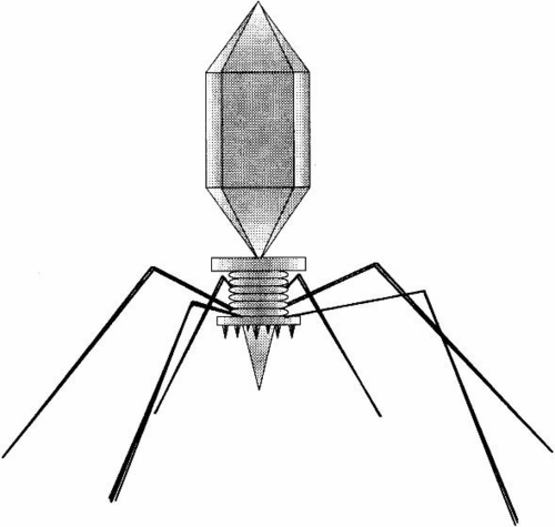
عاثيات متعددة ملتصقة بجدار الخلية البكتيرية بتكبير 200000 مرة
توجد الفيروسات في كل مكان توجد فيه الحياة، وربما وُجدت منذ نشأة الخلايا الحية. [ 35 ] لا يزال أصل الفيروسات غير واضح لأنها لا تُشكّل أحافير، لذا استُخدمت التقنيات الجزيئية لمقارنة الحمض النووي (DNA) أو الحمض النووي الريبي (RNA) للفيروسات، وهي وسيلة مفيدة لدراسة كيفية نشأتها. [ 36 ]
باتت الفيروسات الآن معروفة بأنها قديمة ولها أصول تسبق تفرع الحياة إلى المجالات الثلاثة . [ 37 ]
تتباين الآراء حول ما إذا كانت الفيروسات شكلاً من أشكال الحياة أم مجرد تراكيب عضوية تتفاعل مع الكائنات الحية. [ 34 ] يعتبرها البعض شكلاً من أشكال الحياة، لأنها تحمل مادة وراثية، وتتكاثر عن طريق تكوين نسخ متعددة من نفسها من خلال التجميع الذاتي، وتتطور عبر الانتقاء الطبيعي . ومع ذلك، فهي تفتقر إلى خصائص أساسية كالبنية الخلوية التي تُعتبر ضرورية عمومًا لتصنيفها ككائنات حية. ولأنها تمتلك بعضًا من هذه الخصائص دون غيرها، فقد وُصفت الفيروسات بأنها كائنات مُتكاثرة [ 34 ] وبأنها "كائنات على حافة الحياة". [ 33 ]
العاثيات ، التي تُعرف غالبًا باسم الفيروسات ، هي فيروسات تتطفل على البكتيريا والعتائق. تتطفل العاثيات البحرية على البكتيريا والعتائق البحرية، مثل البكتيريا الزرقاء . [ 38 ] وهي مجموعة شائعة ومتنوعة من الفيروسات، وتُعدّ أكثر الكائنات الحية وفرةً في البيئات البحرية، لأن عوائلها، أي البكتيريا، هي عادةً الكائنات الخلوية المهيمنة عدديًا في البحر. عمومًا، يوجد ما بين مليون إلى عشرة ملايين فيروس في كل ملليلتر من مياه البحر، أي ما يقارب عشرة أضعاف عدد فيروسات الحمض النووي الريبوزي منقوص الأكسجين ثنائي السلسلة مقارنةً بالكائنات الخلوية، [ 39 ] [ 40 ] مع العلم أن تقديرات وفرة الفيروسات في مياه البحر قد تتباين على نطاق واسع. [ 41 ] [ 42 ] لفترة طويلة، بدت العاثيات الذيلية من رتبة Caudovirales مهيمنة على النظم البيئية البحرية من حيث العدد وتنوع الكائنات الحية. [ 38 ] مع ذلك، ونتيجةً لأبحاثٍ أحدث، يبدو أن الفيروسات غير الذيلية هي السائدة في أعماقٍ ومناطقَ محيطيةٍ متعددة، تليها عائلات Caudovirales من الفيروسات العضلية، والفيروسات القدمية، والفيروسات السيفية. [ 43 ] ومن المعروف أيضًا أن العاثيات التي تنتمي إلى عائلات: Corticoviridae ، [ 44 ] وInoviridae ، [ 45 ] و Microviridae ، [ 46 ] و Autolykiviridae [ 47 ] [ 48 ] [ 49 ] [ 50 ] تصيب أنواعًا مختلفةً من البكتيريا البحرية.
توجد أيضًا فيروسات أثرية تتكاثر داخل العتائق : وهي فيروسات ذات حمض نووي مزدوج السلسلة، تتميز بأشكال غير عادية، وأحيانًا فريدة. [ 51 ] [ 52 ] وقد دُرست هذه الفيروسات بتفصيل كبير في العتائق المحبة للحرارة ، ولا سيما رتبتي السلفولوباليس والثيرموبروتياليس . [ 53 ]
دور الفيروسات
تُشكّل الكائنات الدقيقة حوالي 70% من الكتلة الحيوية البحرية. [ 9 ] ويُقدّر أن الفيروسات تقضي على 20% من هذه الكتلة الحيوية يوميًا، وأن عدد الفيروسات في المحيطات يفوق عدد البكتيريا والعتائق بخمسة عشر ضعفًا. تُعدّ الفيروسات العامل الرئيسي المسؤول عن التدمير السريع لتكاثر الطحالب الضارة ، [ 40 ] والتي غالبًا ما تُهلك الكائنات البحرية الأخرى. [ 54 ] ويتناقص عدد الفيروسات في المحيطات كلما ابتعدنا عن الشاطئ وتعمقنا في المياه، حيث يقلّ عدد الكائنات المضيفة. [ 16 ]
تُعدّ الفيروسات وسيلة طبيعية مهمة لنقل الجينات بين الأنواع المختلفة، مما يزيد التنوع الجيني ويدفع عجلة التطور. [ 32 ] يُعتقد أن الفيروسات لعبت دورًا محوريًا في التطور المبكر، قبل تنوع البكتيريا والعتائق وحقيقيات النوى، في زمن السلف المشترك الأخير للحياة على الأرض. [ 55 ] ولا تزال الفيروسات تُشكّل أحد أكبر خزانات التنوع الجيني غير المستكشف على كوكب الأرض. [ 16 ]
يتراوح طول الفيروسات عادةً بين 20 و300 نانومتر. ويُقارن هذا بطول البكتيريا الذي يبدأ من حوالي 400 نانومتر. وهناك أيضًا فيروسات عملاقة ، تُعرف غالبًا باسم الفيروسات العملاقة ، ويبلغ طولها عادةً حوالي 1000 نانومتر (ميكرون واحد). تنتمي جميع الفيروسات العملاقة إلى شعبة الفيروسات الخلوية النووية (NCLDV)، إلى جانب فيروسات الجدري . أكبر هذه الفيروسات المعروفة هو فيروس توبان . اكتُشف هذا الجنس من الفيروسات العملاقة عام 2018 في أعماق المحيط وفي بحيرة صودا، ويمكن أن يصل طوله الإجمالي إلى 2.3 ميكرون. [ 56 ]
أثار اكتشاف الفيروسات العملاقة وتوصيفها لاحقًا جدلًا حول أصولها التطورية. [ 57 ] وتتمثل الفرضيتان الرئيسيتان لأصلها في أنها إما تطورت من فيروسات صغيرة، اكتسبت الحمض النووي من الكائنات المضيفة، أو أنها تطورت من كائنات بالغة التعقيد إلى شكلها الحالي غير القادر على التكاثر ذاتيًا. [ 58 ] كما أن نوع الكائن المعقد الذي ربما انحدرت منه الفيروسات العملاقة هو أيضًا موضوع نقاش. ويُقترح أن نقطة الأصل تمثل في الواقع نطاقًا رابعًا من نطاقات الحياة، [ 59 ] [ 60 ] ولكن تم دحض هذا الاقتراح إلى حد كبير. [ 61 ] [ 62 ]
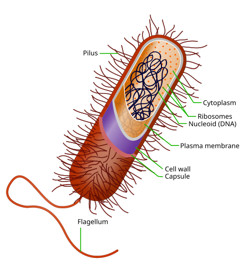
كانت البكتيريا تُصنّف سابقًا ضمن النباتات التي تُشكّل فئة الفطريات المنشقة ، أما الآن فتُصنّف ضمن بدائيات النوى . وعلى عكس خلايا الحيوانات وحقيقيات النوى الأخرى ، لا تحتوي الخلايا البكتيرية على نواة ، ونادرًا ما تحتوي على عضيات محاطة بغشاء . ورغم أن مصطلح البكتيريا كان يشمل تقليديًا جميع بدائيات النوى، فقد تغيّر التصنيف العلمي بعد اكتشاف في تسعينيات القرن الماضي أن بدائيات النوى تتكون من مجموعتين مختلفتين تمامًا من الكائنات الحية التي تطورت من سلف مشترك قديم. تُسمى هاتان المجموعتان التطوريتان بالبكتيريا والعتائق . [ 64 ]
كانت أسلاف البكتيريا الحديثة كائنات دقيقة وحيدة الخلية، تُعدّ أولى أشكال الحياة التي ظهرت على الأرض قبل حوالي 4 مليارات سنة. ولمدة 3 مليارات سنة تقريبًا، كانت معظم الكائنات الحية مجهرية، وكانت البكتيريا والعتائق هي الأشكال السائدة للحياة. [ 65 ] [ 66 ] على الرغم من وجود أحافير بكتيرية ، مثل الستروماتوليت ، إلا أن افتقارها إلى شكل مميز يحول دون استخدامها لدراسة تاريخ تطور البكتيريا، أو لتحديد تاريخ نشأة نوع بكتيري معين. مع ذلك، يمكن استخدام تسلسلات الجينات لإعادة بناء شجرة تطور البكتيريا ، وتشير هذه الدراسات إلى أن البكتيريا انفصلت أولًا عن سلالة العتائق/حقيقيات النوى. [ 67 ] كما شاركت البكتيريا في التباين التطوري الكبير الثاني، وهو تباين العتائق وحقيقيات النوى. هنا، نشأت حقيقيات النوى نتيجة دخول بكتيريا قديمة في علاقات تكافلية داخلية مع أسلاف خلايا حقيقيات النوى، والتي ربما كانت بدورها مرتبطة بالعتائق . [ 68 ] [ 69 ] وقد تضمن ذلك ابتلاع خلايا حقيقية النوى الأولية لمتعايشات من بكتيريا ألفا بروتيو لتكوين إما ميتوكوندريا أو هيدروجينوسومات ، والتي لا تزال موجودة في جميع حقيقيات النوى المعروفة. لاحقًا، ابتلعت بعض حقيقيات النوى التي كانت تحتوي بالفعل على ميتوكوندريا كائنات شبيهة بالبكتيريا الزرقاء. أدى ذلك إلى تكوين البلاستيدات الخضراء في الطحالب والنباتات. كما توجد بعض الطحالب التي نشأت من أحداث تكافلية داخلية لاحقة. هنا، ابتلعت حقيقيات النوى طحالب حقيقية النوى تطورت إلى بلاستيدة من "الجيل الثاني". [ 70 ] [ 71 ] يُعرف هذا بالتكافل الداخلي الثانوي .
تنمو بكتيريا مارينوموناس أركتيكا داخل الجليد البحري في القطب الشمالي في درجات حرارة تحت الصفر
قد تكون بكتيريا Pelagibacter ubique وما يرتبط بها من أكثر الكائنات الحية وفرةً في المحيط، بل يُزعم أنها ربما تكون أكثر أنواع البكتيريا وفرةً في العالم. فهي تُشكّل حوالي 25% من جميع خلايا العوالق الميكروبية ، وفي فصل الصيف قد تُمثّل ما يقارب نصف الخلايا الموجودة في مياه سطح المحيطات المعتدلة. ويُقدّر إجمالي وفرة P. ubique وما يرتبط بها بحوالي 2×10²⁸ ميكروب . [ 73 ] ومع ذلك، فقد نُشر في مجلة Nature في فبراير 2013 خبر اكتشاف العاثية HTVC010P ، التي تُهاجم P. ubique ، و"ربما تكون بالفعل أكثر الكائنات الحية شيوعًا على كوكب الأرض". [ 74 ] [ 75 ]
أكبر أنواع البكتيريا المعروفة، وهي بكتيريا Thiomargarita namibiensis البحرية ، يمكن رؤيتها بالعين المجردة، ويصل حجمها أحيانًا إلى 0.75 ملم (750 ميكرومتر) . [ 76 ] [ 77 ]
العتائق البحرية
كان يُنظر إلى العتائق في البداية على أنها كائنات متطرفة تعيش في بيئات قاسية، مثل العتائق الصفراء المصورة هنا في نبع حار ، ولكن تم العثور عليها منذ ذلك الحين في نطاق أوسع بكثير من الموائل . [ 78 ]
صُنفت العتائق في البداية ضمن البكتيريا ، لكن هذا التصنيف قديم. [ 80 ] تتميز خلايا العتائق بخصائص فريدة تميزها عن نطاقي الحياة الآخرين، البكتيريا وحقيقيات النوى . تُقسم العتائق بدورها إلى عدة شعب معترف بها . ويُعد تصنيفها صعبًا لأن معظمها لم يُعزل في المختبر، وإنما تم اكتشافه فقط من خلال تحليل أحماضها النووية في عينات من بيئتها.
تُعدّ العتائق من الكائنات الحية الدقيقة ذات وفرة كبيرة في المحيطات، وقد تكون العتائق الموجودة في العوالق من أكثر مجموعات الكائنات الحية وفرةً على كوكب الأرض. تُشكّل العتائق جزءًا أساسيًا من الحياة على الأرض، وقد تلعب أدوارًا مهمة في كلٍّ من دورة الكربون ودورة النيتروجين . تُعرف شعبة العتائق باسم Thermoproteota (أو eocytes أو Crenarchaeota)، وهي شعبة من العتائق، ويُعتقد أنها وفيرة جدًا في البيئات البحرية، وتُعدّ من المساهمين الرئيسيين في تثبيت الكربون. [ 82 ]
يمكن تصنيف جميع الكائنات الحية إلى بدائيات النوى وحقيقيات النوى . نشأت الحياة من بدائيات النوى وحيدة الخلية ، ثم تطورت لاحقًا إلى حقيقيات النوى الأكثر تعقيدًا. وعلى عكس خلايا بدائيات النوى، تتميز خلايا حقيقيات النوى بتنظيمها العالي. تشمل بدائيات النوى البكتيريا والعتائق، بينما تشمل حقيقيات النوى أشكال الحياة الأخرى - الطلائعيات والنباتات والفطريات والحيوانات. عادةً ما تكون الطلائعيات وحيدة الخلية، بينما تكون النباتات والفطريات والحيوانات متعددة الخلايا .
الطلائعيات هي كائنات حقيقية النواة لا يمكن تصنيفها ضمن النباتات أو الفطريات أو الحيوانات. وهي عادةً وحيدة الخلية ومجهرية. نشأت الحياة من بدائيات النوى وحيدة الخلية (البكتيريا والعتائق) ثم تطورت لاحقًا إلى حقيقيات نوى أكثر تعقيدًا . حقيقيات النوى هي أشكال الحياة الأكثر تطورًا والمعروفة بالنباتات والحيوانات والفطريات والطلائعيات. شاع استخدام مصطلح "الطلائعيات" تاريخيًا كمصطلح مبسط للكائنات حقيقية النواة التي لا يمكن تصنيفها بدقة ضمن النباتات أو الحيوانات أو الفطريات. وهي ليست جزءًا من علم التصنيف التفرعي الحديث، لأنها شبه عرقية (تفتقر إلى سلف مشترك).
عن طريق النمط الغذائي
يمكن تقسيم الطلائعيات بشكل عام إلى أربع مجموعات اعتمادًا على ما إذا كان نظامها الغذائي نباتيًا أو حيوانيًا أو فطريًا، [ 85 ] أو مزيجًا من هذه الأنظمة. [ 86 ]
تُعدّ الطلائعيات كائنات حية شديدة التنوع، تُصنّف حاليًا ضمن 18 شعبة، إلا أن تصنيفها ليس بالأمر السهل. [ 89 ] [ 90 ] وقد أظهرت الدراسات وجود تنوع كبير في الطلائعيات في المحيطات، وفتحات أعماق البحار، ورواسب الأنهار، مما يشير إلى وجود عدد كبير من المجتمعات الميكروبية حقيقية النواة لم يتم اكتشافها بعد. [ 91 ] [ 92 ] وقد أُجريت أبحاث قليلة على الطلائعيات المختلطة التغذية ، إلا أن الدراسات الحديثة في البيئات البحرية وجدت أن هذه الطلائعيات تُساهم بجزء كبير من الكتلة الحيوية للطلائعيات . [ 87 ] وبما أن الطلائعيات من حقيقيات النوى، فإنها تحتوي داخل خلاياها على نواة واحدة على الأقل ، بالإضافة إلى عضيات مثل الميتوكوندريا وجهاز جولجي . وتتكاثر الطلائعيات لا جنسيًا، ولكنها قادرة على التكاثر بسرعة من خلال الانقسام المتساوي أو التجزؤ .
الطلائعيات وحيدة الخلية والمجهرية
تُعد الدياتومات مجموعة رئيسية من الطحالب، حيث تُنتج حوالي 20% من إنتاج الأكسجين العالمي. [ 93 ]
تتميز الدياتومات بجدران خلوية تشبه الزجاج مصنوعة من السيليكا وتسمى الصدف . [ 94 ]
هيكل دياتوم أحفوري يعود تاريخه إلى ما بين 32 و 40 مليون سنة
على عكس خلايا بدائيات النوى، تتميز خلايا حقيقيات النوى بتنظيمها العالي. فالنباتات والحيوانات والفطريات عادةً ما تكون متعددة الخلايا ، وغالبًا ما تكون مرئية بالعين المجردة . أما معظم الطلائعيات فهي وحيدة الخلية ومجهرية. ولكن هناك استثناءات. فبعض الطلائعيات البحرية وحيدة الخلية مرئية بالعين المجردة. كما أن بعض أنواع العفن الغروي البحري لها دورات حياة فريدة تتضمن التحول بين الأشكال وحيدة الخلية، والمستعمرة ، ومتعددة الخلايا. [ 95 ] وهناك طلائعيات بحرية أخرى ليست وحيدة الخلية ولا مجهرية، مثل الأعشاب البحرية .
وُصفت الطلائعيات بأنها مجموعة تصنيفية متنوعة تضم كائنات لا تنتمي إلى أي من الممالك البيولوجية الرئيسية . [ 98 ] يفضل بعض الباحثين المعاصرين استبعاد الكائنات متعددة الخلايا من التعريف التقليدي للطلائعيات، ويقتصر تعريفها على الكائنات وحيدة الخلية. [ 99 ] [ 100 ] هذا التعريف الأكثر تحديدًا يستبعد العديد من الطحالب البنية ، والطحالب الحمراء والخضراء متعددة الخلايا ، والعفن الغروي . [ 101 ]
عن طريق الحركة
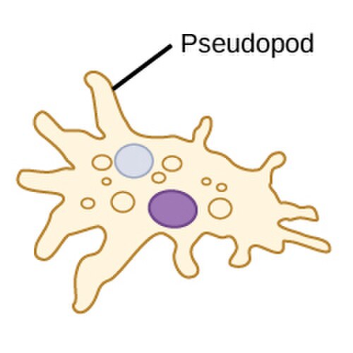
هناك طريقة أخرى لتصنيف الطلائعيات وهي وفقًا لطريقة حركتها. فالعديد من الطلائعيات وحيدة الخلية، وخاصة الأوليات، متحركة وتستطيع توليد الحركة باستخدام الأسواط أو الأهداب أو الأقدام الكاذبة . تُسمى الخلايا التي تستخدم الأسواط للحركة عادةً بالخلايا السوطية ، والخلايا التي تستخدم الأهداب عادةً بالخلايا الهدبية ، والخلايا التي تستخدم الأقدام الكاذبة عادةً بالأميبا أو الأميبات . أما الطلائعيات الأخرى فهي غير متحركة ، وبالتالي لا تمتلك آلية حركة.
السوط (كلمة لاتينية تعني السوط ) هو زائدة تشبه السوط تبرز من جسم الخلية لدى بعض الطلائعيات (وكذلك بعض البكتيريا). تستخدم السوطيات من سوط واحد إلى عدة أسواط للحركة، وأحيانًا كعضية للتغذية والإحساس .
تشمل السوطيات البكتيريا والطلائعيات. يستخدم نموذج المحرك الدوراني الذي تستخدمه البكتيريا بروتونات التدرج الكهروكيميائي لتحريك أسواطها. يتولد عزم الدوران في أسواط البكتيريا بواسطة جزيئات تنقل البروتونات حول قاعدة السوط. وينشأ اتجاه دوران الأسواط في البكتيريا من شغل قنوات البروتونات على طول محيط محرك السوط. [ 107 ]
تمتلك الهدبيات عادةً مئات إلى آلاف الأهداب المتراصة بكثافة في صفوف. أثناء الحركة، يتشوه الهدب الواحد باستخدام ضربة قوة عالية الاحتكاك تليها ضربة استعادة منخفضة الاحتكاك. ونظرًا لوجود أهداب متعددة متراصة على الكائن الحي الواحد، فإنها تُظهر سلوكًا جماعيًا بإيقاع متزامن . وهذا يعني أن تشوه هدب واحد يكون متزامنًا مع تشوه الهدب المجاور له، مما يُسبب موجات تشوه تنتشر على طول سطح الكائن الحي. هذه الموجات المنتشرة من الأهداب هي ما يسمح للكائن الحي باستخدام الأهداب بطريقة منسقة للحركة. ومن الأمثلة النموذجية على الكائنات الحية الدقيقة الهدبية البراميسيوم ، وهو كائن أولي وحيد الخلية مُهدب مغطى بآلاف الأهداب. تسمح الأهداب التي تنبض معًا للبراميسيوم بالتحرك عبر الماء بسرعة تصل إلى 500 ميكرومتر في الثانية. [ 108 ]
يحتوي المليلتر الواحد من مياه البحر على ما يقارب 10³ إلى 10⁴ خلية فطرية. [ 119 ] ويزداد هذا العدد في النظم البيئية الساحلية ومصبات الأنهار نتيجةً للجريان السطحي المغذي من المجتمعات الأرضية. ويُلاحظ تنوع أكبر في العوالق الفطرية حول السواحل وفي المياه السطحية حتى عمق 1000 متر، مع توزيع رأسي يعتمد على وفرة العوالق النباتية . [ 120 ] [ 121 ] ويتغير هذا التوزيع بين الفصول نتيجةً لتغيرات توافر المغذيات. [ 122 ] وتعيش الفطريات البحرية في بيئة تعاني من نقص الأكسجين بشكل مستمر، ولذلك تعتمد على انتشار الأكسجين بفعل الاضطراب والأكسجين الناتج عن الكائنات الحية التي تقوم بعملية التمثيل الضوئي . [ 123 ]
يمكن تصنيف الفطريات البحرية على النحو التالي: [ 123 ]
الأشنات هي علاقات تكافلية بين فطر، عادةً ما يكون من الفطريات الزقية ، وطحلب أو بكتيريا زرقاء . توجد أنواع عديدة من الأشنات في البيئات البحرية. [ 124 ] ويوجد عدد أكبر منها في منطقة الرذاذ ، حيث تشغل مناطق رأسية مختلفة تبعًا لمدى تحملها للغمر. [ 125 ] تعيش بعض الأشنات لفترات طويلة؛ فقد تم تحديد عمر أحد الأنواع بـ 8600 عام. [ 126 ] ومع ذلك، يصعب قياس عمرها لأن تعريف الأشنة الواحدة ليس دقيقًا. [ 127 ] تنمو الأشنات عن طريق التكاثر الخضري، حيث تنفصل أجزاء منها، وقد تُعتبر أو لا تُعتبر من نفس النوع، كما يمكن أن تندمج أشنتان مختلفتان في العمر، مما يثير التساؤل حول ما إذا كانتا من نفس النوع. [ 127 ]
يُلحق حلزون البحر Littoraria irrorata الضرر بنباتات Spartina في المستنقعات البحرية التي يعيش فيها، مما يُتيح لأبواغ الفطريات الزقية التي تعيش بين المد والجزر استعمار النبات. ثم يتغذى الحلزون على نمو الفطريات بدلاً من العشب نفسه. [ 128 ]
تشير السجلات الأحفورية إلى أن الفطريات تعود إلى أواخر حقبة البروتيروزويك، أي قبل 900 إلى 570 مليون سنة. وقد اكتُشفت في الصين أحافير لأشنات بحرية عمرها 600 مليون سنة. [ 129 ] ويُفترض أن العوالق الفطرية تطورت من الفطريات الأرضية، على الأرجح في حقبة الباليوزويك (قبل 390 مليون سنة). [ 130 ]
صورة مركبة توضح التوزيع العالمي لعملية التمثيل الضوئي، بما في ذلك العوالق النباتية في المحيطات والنباتات البرية . يشير اللون الأحمر الداكن والأزرق المخضر إلى مناطق ذات نشاط تمثيلي ضوئي عالٍ في المحيط وعلى اليابسة، على التوالي.
المنتجون الأوليون هم الكائنات ذاتية التغذية التي تصنع غذائها بنفسها بدلاً من التهام كائنات أخرى. وهذا يعني أن المنتجين الأوليين يمثلون نقطة البداية في السلسلة الغذائية للكائنات غيرية التغذية التي تتغذى على كائنات أخرى. بعض المنتجين الأوليين البحريين عبارة عن بكتيريا وعتائق متخصصة، وهي كائنات كيميائية التغذية ، تصنع غذائها بنفسها من خلال التجمع حول الفتحات الحرارية المائية والينابيع الباردة باستخدام عملية التخليق الكيميائي . مع ذلك، فإن معظم الإنتاج الأولي البحري يأتي من كائنات تستخدم عملية التمثيل الضوئي على ثاني أكسيد الكربون المذاب في الماء. تستخدم هذه العملية طاقة ضوء الشمس لتحويل الماء وثاني أكسيد الكربون [ 133 ] : 186-187 إلى سكريات يمكن استخدامها كمصدر للطاقة الكيميائية والجزيئات العضوية التي تدخل في تركيب الخلايا. [ 133 ] : 1242. تكمن أهمية المنتجين الأوليين البحريين في كونهم أساس الحياة الحيوانية البحرية، حيث يوفرون معظم الأكسجين والغذاء اللازمين لتزويد الكائنات الأخرى بالطاقة الكيميائية التي تحتاجها للبقاء.
كانت البكتيريا الزرقاء أول الكائنات الحية التي طورت القدرة على تحويل ضوء الشمس إلى طاقة كيميائية. وهي تشكل شعبة (قسمًا) من البكتيريا تتراوح بين وحيدة الخلية والخيطية ، وتشمل أنواعًا مستعمرة . وتوجد في كل مكان تقريبًا على سطح الأرض: في التربة الرطبة، وفي كل من البيئات المائية العذبة والمالحة، وحتى على صخور القطب الجنوبي. [ 136 ] وعلى وجه الخصوص، توجد بعض الأنواع كخلايا طافية في المحيط، ولذلك كانت من أوائل العوالق النباتية .
كانت البكتيريا الزرقاء المحيطية أول الكائنات المنتجة الأولية التي استخدمت عملية التمثيل الضوئي، وذلك قبل حوالي 2.3 مليار سنة. [ 137 ] [ 138 ] وقد أدى إطلاق الأكسجين الجزيئي من قِبل البكتيريا الزرقاء كمنتج ثانوي لعملية التمثيل الضوئي إلى تغييرات عالمية في بيئة الأرض. ولأن الأكسجين كان سامًا لمعظم أشكال الحياة على الأرض في ذلك الوقت، فقد أدى ذلك إلى انقراض شبه كامل للكائنات الحية التي لا تتحمل الأكسجين ، وهو تغيير جذري أعاد توجيه مسار تطور الأنواع الرئيسية من الحيوانات والنباتات. [ 139 ]
تُشكّل بكتيريا البروكلوروكوكس الزرقاء البحرية الدقيقة (0.6 ميكرومتر ) ، التي اكتُشفت عام 1986، جزءًا هامًا من قاعدة السلسلة الغذائية في المحيطات ، وتُساهم بشكل كبير في عملية التمثيل الضوئي في المحيطات المفتوحة [ 140 ] ، وتُقدّر نسبتها بنحو 20% من الأكسجين في الغلاف الجوي للأرض. [ 141 ] وربما تكون أكثر الأجناس وفرةً على وجه الأرض: إذ قد يحتوي ملليلتر واحد من مياه البحر السطحية على 100,000 خلية أو أكثر. [ 142 ]
في الأصل، اعتقد علماء الأحياء أن البكتيريا الزرقاء هي طحالب، وأطلقوا عليها اسم "الطحالب الخضراء المزرقة". أما الرأي السائد حاليًا فهو أن البكتيريا الزرقاء هي بكتيريا، وبالتالي فهي لا تنتمي حتى إلى نفس مملكة الطحالب. وتستبعد معظم المراجع العلمية جميع بدائيات النوى ، وبالتالي البكتيريا الزرقاء، من تعريف الطحالب. [ 143 ] [ 144 ]
تُعدّ الدياتومات والدينوفلاجيلات مكوناتٍ مهمةً للطحالب البحرية، ولها أقسامٌ خاصةٌ بها أدناه. أما اليوجلينات فهي شعبةٌ من الكائنات السوطية وحيدة الخلية، ولا يوجد منها سوى عددٍ قليلٍ من الكائنات البحرية.
ليست كل الطحالب مجهرية. فالطحالب الخضراء والحمراء والبنية لها أشكال متعددة الخلايا مرئية بالعين المجردة، وهي التي تُشكل الأعشاب البحرية المألوفة . تضم الطحالب الخضراء ، وهي مجموعة غير رسمية، حوالي 8000 نوع مُعترف به. [ 145 ] تعيش العديد من الأنواع معظم حياتها كخلايا منفردة أو خيطية الشكل، بينما تُشكل أنواع أخرى مستعمرات تتكون من سلاسل طويلة من الخلايا، أو هي أعشاب بحرية مرئية بالعين المجردة ومتمايزة للغاية. أما الطحالب الحمراء ، وهي شعبة (مُختلف عليها) تضم حوالي 7000 نوع مُعترف به، [ 146 ] معظمها متعدد الخلايا ، وتشمل العديد من الأعشاب البحرية البارزة. [ 146 ] [ 147 ] تُشكل الطحالب البنية فئة تضم حوالي 2000 نوع مُعترف به، [ 148 ] معظمها متعدد الخلايا، وتشمل العديد من الأعشاب البحرية مثل عشب البحر . على عكس النباتات الراقية، تفتقر الطحالب إلى الجذور والسيقان والأوراق. ويمكن تصنيفها حسب الحجم إلى طحالب دقيقة وطحالب كبيرة .
الطحالب الدقيقة هي أنواع مجهرية من الطحالب، لا تُرى بالعين المجردة. وهي في الغالب كائنات وحيدة الخلية ، تعيش منفردة أو في سلاسل أو مجموعات، مع أن بعضها متعدد الخلايا . تُعدّ الطحالب الدقيقة مكونات أساسية للطلائعيات البحرية المذكورة سابقًا ، وكذلك للعوالق النباتية المذكورة لاحقًا . وهي شديدة التنوع ، إذ يُقدّر عدد أنواعها بين 200,000 و800,000 نوع، وُصِف منها حوالي 50,000 نوع. [ 149 ] وتتراوح أحجامها، بحسب النوع، من بضعة ميكرومترات إلى بضع مئات من الميكرومترات. وهي مُتكيّفة بشكل خاص مع بيئة تهيمن عليها قوى اللزوجة.
الكائنات وحيدة الخلية عادةً ما تكون مجهرية، ولكن هناك استثناءات. فمثلاً، تُعدّ طحالب "كأس حورية البحر" ، وهي جنس من الطحالب الخضراء شبه الاستوائية ، كائناً وحيد الخلية، إلا أنها كبيرة الحجم ومعقدة الشكل بشكل ملحوظ، إذ تحتوي على نواة واحدة كبيرة، مما يجعلها كائناً نموذجياً لدراسة بيولوجيا الخلية . [ 150 ] أما طحلب " كوليربا تاكسيفوليا "، وهو طحلب آخر وحيد الخلية ، فيشبه في مظهره النباتات الوعائية، بما في ذلك "الأوراق" المرتبة بدقة على سيقان تشبه السرخس. وقد أدى التكاثر الانتقائي في أحواض السمك لإنتاج سلالات أكثر مقاومة إلى إطلاقها عن طريق الخطأ في البحر الأبيض المتوسط، حيث أصبحت نوعاً غازياً يُعرف شعبياً باسم " الطحالب القاتلة " . [ 151 ]
الكلاميدوموناس غلوبوزا ، طحلب أخضر وحيد الخلية ذو سوطين يظهران بالكاد في أسفل اليسار
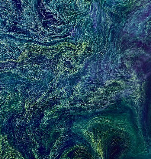
العوالق هي كائنات حية تنجرف أو تطفو ولا تستطيع السباحة بفعالية ضد التيار، وتشمل كائنات حية من معظم مجالات الحياة: البكتيريا ، والعتائق ، والطحالب ، والبروتوزوا ، والحيوانات .تمثل الألوان الأحمر والبرتقالي والأصفر والأخضر المناطق التي تكثر فيها الطحالب. أما المناطق الزرقاء فتمثل المناطق الفقيرة بالمغذيات حيث يوجد العوالق النباتية بتركيزات أقل.
العوالق (من اليونانية وتعني " الهائمون ") هي مجموعة متنوعة من الكائنات الحية التي تعيش في عمود الماء في المسطحات المائية الكبيرة، لكنها لا تستطيع السباحة عكس التيار. ونتيجة لذلك، فإنها تتجول أو تنجرف مع التيارات. [ 153 ] تُعرَّف العوالق من خلال بيئتها المتخصصة ، وليس من خلال أي تصنيف تطوري أو تصنيفي . وهي مصدر غذائي أساسي للعديد من الحيوانات البحرية، من الأسماك الصغيرة إلى الحيتان . يمكن تقسيم العوالق إلى مكون نباتي ومكون حيواني.
العوالق النباتية
تُصدر العوالق النباتية، مثل مستعمرة Chaetoceros socialis هذه ، ضوءًا أحمرًا فلوريًا طبيعيًا يُبدد الطاقة الشمسية الزائدة التي لا تستطيع استهلاكها من خلال عملية التمثيل الضوئي. ويمكن للأقمار الصناعية رصد هذا التوهج كمؤشر على كفاءة عملية التمثيل الضوئي في العوالق النباتية في المحيط. [ 154 ] [ 155 ]
العوالق النباتية هي المكونات الشبيهة بالنباتات في مجتمع العوالق (كلمة "فيتو" مشتقة من الكلمة اليونانية التي تعني نبات ). وهي ذاتية التغذية، أي أنها تنتج غذائها بنفسها ولا تحتاج إلى استهلاك كائنات حية أخرى.
تؤدي العوالق النباتية ثلاث وظائف حيوية: فهي تُنتج ما يقارب نصف الأكسجين الجوي في العالم، وتُنظم مستويات ثاني أكسيد الكربون في المحيطات والغلاف الجوي، وتُشكل قاعدة السلسلة الغذائية البحرية . وعندما تكون الظروف مواتية، قد تحدث تكاثرات هائلة للطحالب العوالق النباتية في المياه السطحية. وتُعد العوالق النباتية من الكائنات الحية سريعة النمو، إذ يمكنها مضاعفة أعدادها يوميًا. وقد تُصبح هذه التكاثرات سامة وتُستنزف الأكسجين من الماء. ومع ذلك، عادةً ما تبقى أعداد العوالق النباتية تحت السيطرة من خلال استهلاكها للعناصر الغذائية المتاحة، ومن خلال رعيها للعوالق الحيوانية. [ 156 ]
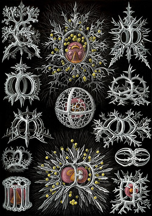
رسومات من تصميم هايكل عام 1904 (انقر للحصول على التفاصيل)
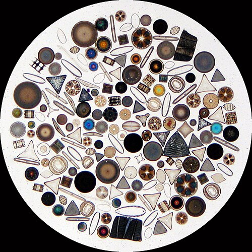
تشكل الدياتومات شعبة (مختلف عليها) تضم حوالي 100,000 نوع معترف به من الطحالب وحيدة الخلية في الغالب. وتنتج الدياتومات حوالي 20% من الأكسجين المنتج على كوكب الأرض كل عام، [ 93 ] وتمتص أكثر من 6.7 مليار طن متري من السيليكون كل عام من المياه التي تعيش فيها، [ 159 ] وتساهم بنحو نصف المواد العضوية الموجودة في المحيطات.
تمتلك الدياتومات غلافًا من السيليكا ( فروستول ) ذو تناظر شعاعي (مركزي) أو ثنائي (ريشي)
تُحاط الدياتومات بأغلفة واقية من السيليكا (الزجاج) تُسمى الصدفة . تتكون كل صدفة من جزأين متشابكين مغطيين بثقوب دقيقة تتبادل من خلالها الدياتومات المغذيات والفضلات. [ 156 ] تنجرف صدفات الدياتومات الميتة إلى قاع المحيط، حيث يمكن أن تتراكم على مدى ملايين السنين ليصل عمقها إلى نصف ميل . [ 160 ]
غلاف متسيليكي لدياتوم ريشي الشكل بنصفين متداخلين
Guinardia delicatula ، وهي دياتوم مسؤولة عن ازدهار الطحالب في بحر الشمال والقناة الإنجليزية [ 161 ].
دياتوم أحفوري
يوجد أكثر من 100,000 نوع من الدياتومات التي تمثل 50% من الإنتاج الأولي للمحيطات
الكوكوليثوفورات كائنات أولية وحيدة الخلية دقيقة الحجم، تقوم بعملية التمثيل الضوئي، ولها سوطان للحركة. معظمها محمي بقشرة مغطاة بصفائح أو حراشف دائرية مزخرفة تُسمى الكوكوليثات . تتكون الكوكوليثات من كربونات الكالسيوم. تُعد قشور الكالسيت مهمة لدورة الكربون البحرية. [ 163 ] يُشتق مصطلح الكوكوليثوفور من الكلمة اليونانية التي تعني حجرًا يحمل بذرة ، في إشارة إلى صغر حجمها وأحجار الكوكوليث التي تحملها. في ظل الظروف المناسبة، تتكاثر هذه الكائنات، مثل العوالق النباتية الأخرى، ويمكنها أن تُحوّل المحيط إلى اللون الأبيض الحليبي. [ 164 ]
ازدهار طحالب إيميليانيا هوكسلي قبالة الساحل الجنوبي لإنجلترا
أحفورة منقرضة
الرودوبسين الميكروبي
نموذج لآلية توليد الطاقة في البكتيريا البحرية
(1) عندما يسقط ضوء الشمس على جزيء الرودوبسين (2) فإنه يغير شكله بحيث يتم طرد بروتون من الخلية (3) ويتسبب الجهد الكيميائي في تدفق البروتون عائدًا إلى الخلية (4) وبالتالي توليد الطاقة (5) على شكل أدينوسين ثلاثي الفوسفات . [ 165 ]
يعتمد التمثيل الغذائي الضوئي على أحد ثلاثة أصباغ محولة للطاقة: الكلوروفيل ، والبكتيريوكلوروفيل ، والريتينال . والريتينال هو الصباغ الموجود في الرودوبسينات . وقد كُتب عن أهمية الكلوروفيل في تحويل الطاقة الضوئية لعقود، لكن دراسة التمثيل الغذائي الضوئي القائم على أصباغ الريتينال بدأت للتو. [ 166 ]
في عام 2000، حقق فريق من علماء الأحياء الدقيقة بقيادة إدوارد ديلونغ اكتشافًا بالغ الأهمية في فهم دورات الكربون والطاقة في البيئة البحرية. فقد اكتشفوا جينًا في عدة أنواع من البكتيريا [ 168 ] [ 169 ] مسؤولًا عن إنتاج بروتين الرودوبسين ، وهو بروتين لم يكن معروفًا من قبل في البكتيريا. هذه البروتينات الموجودة في أغشية الخلايا قادرة على تحويل الطاقة الضوئية إلى طاقة كيميائية حيوية نتيجة لتغير في بنية جزيء الرودوبسين عند تعرضه لأشعة الشمس، مما يؤدي إلى ضخ بروتون من الداخل إلى الخارج، ثم تدفق البروتون إلى الداخل لتوليد الطاقة. [ 170 ] وقد وُجدت أنواع الرودوبسين الشبيهة بتلك الموجودة في العتائق لاحقًا في مختلف التصنيفات، بما في ذلك الطلائعيات والبكتيريا والعتائق، على الرغم من ندرتها في الكائنات الحية المعقدة متعددة الخلايا . [ 171 ] [ 172 ] [ 173 ]
أظهرت الأبحاث التي أُجريت عام 2019 أن هذه "البكتيريا المُستَغِلّة لأشعة الشمس" أكثر انتشارًا مما كان يُعتقد سابقًا، وقد تُغيّر كيفية تأثر المحيطات بالاحتباس الحراري. "تُخالف هذه النتائج التفسير التقليدي لعلم البيئة البحرية الوارد في الكتب الدراسية، والذي ينص على أن الكلوروفيل الموجود في الطحالب يمتصّ تقريبًا كل ضوء الشمس في المحيط. في المقابل، تعمل البكتيريا المُجهّزة بالرودوبسين كسيارات هجينة، إذ تستمد طاقتها من المواد العضوية عند توفّرها - كما هو الحال مع معظم البكتيريا - ومن ضوء الشمس عندما تكون العناصر الغذائية شحيحة." [ 174 ] [ 166 ]
خلال ثلاثينيات القرن العشرين، وجد ألفريد سي. ريدفيلد أوجه تشابه بين تركيب العناصر في العوالق النباتية والمغذيات الذائبة الرئيسية في أعماق المحيط. [ 177 ] اقترح ريدفيلد أن نسبة الكربون إلى النيتروجين إلى الفوسفور (106:16:1) في المحيط تخضع لاحتياجات العوالق النباتية، حيث تُطلق العوالق النباتية النيتروجين والفوسفور لاحقًا أثناء إعادة تمعدنها. تُعرف هذه النسبة بنسبة ريدفيلد ، وتُستخدم كمبدأ أساسي في وصف قياس العناصر في مياه البحر وتطور العوالق النباتية. [ 178 ]
مع ذلك، فإن نسبة ريدفيلد ليست قيمة ثابتة، بل تتغير تبعًا لعوامل مثل خط العرض الجغرافي. [ 179 ] وبناءً على توزيع الموارد، يمكن تصنيف العوالق النباتية إلى ثلاث استراتيجيات نمو مختلفة: البقاء، والتكاثر السريع، والنمو العام. تتميز العوالق النباتية البقاءية بنسبة نيتروجين إلى فسفور عالية (>30)، وتحتوي على وفرة من آليات اكتساب الموارد اللازمة لاستدامة نموها في ظل ندرة الموارد. أما العوالق النباتية التكاثرية، فتتميز بنسبة نيتروجين إلى فسفور منخفضة (<10)، وتحتوي على نسبة عالية من آليات النمو، وهي متكيفة مع النمو الأسي. بينما تتميز العوالق النباتية العامة بنسبة نيتروجين إلى فسفور مماثلة لنسبة ريدفيلد، وتحتوي على آليات اكتساب موارد ونمو متساوية نسبيًا. [ 178 ]
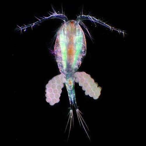
العوالق الحيوانية هي المكون الحيواني للمجتمع العوالقي (كلمة "حيوان" مشتقة من الكلمة اليونانية التي تعني حيوان ). وهي كائنات غيرية التغذية، أي أنها لا تستطيع إنتاج غذائها بنفسها، بل تتغذى على النباتات أو الحيوانات الأخرى. وبالتحديد، هذا يعني أنها تتغذى على العوالق النباتية.
رسومات من تصميم هايكل عام 1904 (انقر للحصول على التفاصيل)
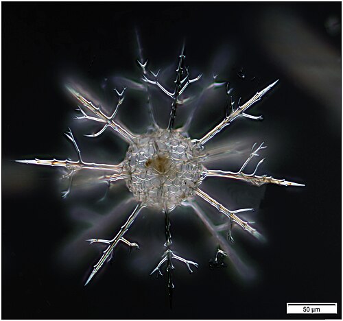
الراديولاريا كائنات أولية مفترسة وحيدة الخلية ، محاطة بأصداف كروية معقدة، مصنوعة عادةً من السيليكا ومثقوبة. اسمها مشتق من الكلمة اللاتينية التي تعني "نصف القطر". تصطاد فرائسها عن طريق مدّ أجزاء من جسمها عبر هذه الثقوب. وكما هو الحال مع قشور السيليكا للدياتومات، يمكن لأصداف الراديولاريا أن تغرق إلى قاع المحيط عند موتها، وتُحفظ كجزء من رواسب المحيط . توفر هذه البقايا، باعتبارها أحافير دقيقة ، معلومات قيّمة عن ظروف المحيطات في الماضي. [ 185 ]
مثل الدياتومات، تأتي الشعاعيات بأشكال عديدة
وكما هو الحال مع الدياتومات، فإن أصداف الشعاعيات تتكون عادة من السيليكات.
محاكاة الحاسوب لأنماط تورينج على الكرة تحاكي بشكل وثيق بعض أنماط أصداف الراديولاريا [ 186 ]
المنخربات
أشكال المثقبات
رسومات من تصميم هايكل عام 1904 (انقر للحصول على التفاصيل)
مثل الشعاعيات، تُعدّ المنخربات ( أو المنخربات اختصارًا) كائنات أولية مفترسة وحيدة الخلية، محمية أيضًا بأصداف مثقوبة. اسمها مشتق من الكلمة اللاتينية التي تعني "حاملات الثقوب". أصدافها، التي تُسمى غالبًا بالهياكل ، مُقسّمة إلى حجرات (تضيف المنخربات المزيد من الحجرات مع نموها). تتكون الأصداف عادةً من الكالسيت، ولكنها تتكون أحيانًا من جزيئات رسوبية مُتكتلة أو الكيتون ، ونادرًا من السيليكا. معظم المنخربات قاعية، ولكن حوالي 40 نوعًا منها عوالق. [ 187 ] تخضع هذه الكائنات لدراسات واسعة النطاق، ولديها سجلات أحفورية موثقة جيدًا تسمح للعلماء باستنتاج الكثير عن البيئات والمناخات الماضية. [ 185 ]
المنخربات
...يمكن أن يحتوي على أكثر من نواة واحدة
...والأعمدة الدفاعية
تُعدّ المنخربات من الطلائعيات وحيدة الخلية المهمة في العوالق الحيوانية ، وتتميز باختبارات الكالسيوم.
Phaeocystis is an important algal genus found as part of the marine phytoplankton around the world. It has a polymorphic life cycle, ranging from free-living cells to large colonies.[197] It has the ability to form floating colonies, where hundreds of cells are embedded in a gel matrix, which can increase massively in size during blooms.[198] As a result, Phaeocystis is an important contributor to the marine carbon[199] and sulfur cycles.[200]Phaeocystis species are endosymbionts to acantharian radiolarians.[201][202]
Mixotrophic plankton that combine phototrophy and heterotrophy – table based on Stoecker et al., 2017[193]
Called constitutive mixotrophs by Mitra et al., 2016.[203] Phytoplankton that eat: photosynthetic protists with inherited plastids and the capacity to ingest prey.
Called nonconstitutive mixotrophs by Mitra et al., 2016.[203] Zooplankton that are photosynthetic: microzooplankton or metazoan zooplankton that acquire phototrophy through chloroplast retentiona or maintenance of algal endosymbionts.
Generalists
Protists that retain chloroplasts and rarely other organelles from many algal taxa
Dinoflagellates are part of the algae group, and form a phylum of unicellular flagellates with about 2,000 marine species.[204] The name comes from the Greek "dinos" meaning whirling and the Latin "flagellum" meaning a whip or lash. This refers to the two whip-like attachments (flagella) used for forward movement. Most dinoflagellates are protected with red-brown, cellulose armour. Like other phytoplankton, dinoflagellates are r-strategists which under right conditions can bloom and create red tides. Excavates may be the most basal flagellate lineage.[102]
By trophic orientation dinoflagellates cannot be uniformly categorized. Some dinoflagellates are known to be photosynthetic, but a large fraction of these are in fact mixotrophic, combining photosynthesis with ingestion of prey (phagotrophy).[205] Some species are endosymbionts of marine animals and other protists, and play an important part in the biology of coral reefs. Others predate other protozoa, and a few forms are parasitic. Many dinoflagellates are mixotrophic and could also be classified as phytoplankton.
The toxic dinoflagellate Dinophysis acuta acquire chloroplasts from its prey. "It cannot catch the cryptophytes by itself, and instead relies on ingesting ciliates such as the red Myrionecta rubra, which sequester their chloroplasts from a specific cryptophyte clade (Geminigera/Plagioselmis/Teleaulax)".[193]
Gyrodinium, one of the few naked dinoflagellates which lack armour
The dinoflagellate Protoperidinium extrudes a large feeding veil to capture prey
Nassellarian radiolarians can be in symbiosis with dinoflagellates
The dinoflagellate Dinophysis acuta
A surf wave at night sparkles with blue light due to the presence of a bioluminescent dinoflagellate, such as Lingulodinium polyedrum
Traditionally dinoflagellates have been presented as armoured or unarmoured
Dinoflagellates often live in symbiosis with other organisms. Many nassellarian radiolarians house dinoflagellatesymbionts within their tests.[207] The nassellarian provides ammonium and carbon dioxide for the dinoflagellate, while the dinoflagellate provides the nassellarian with a mucous membrane useful for hunting and protection against harmful invaders.[208] There is evidence from DNA analysis that dinoflagellate symbiosis with radiolarians evolved independently from other dinoflagellate symbioses, such as with foraminifera.[209]
Some dinoflagellates are bioluminescent. At night, ocean water can light up internally and sparkle with blue light because of these dinoflagellates.[210][211] Bioluminescent dinoflagellates possess scintillons, individual cytoplasmic bodies which contain dinoflagellate luciferase, the main enzyme involved in the luminescence. The luminescence, sometimes called the phosphorescence of the sea, occurs as brief (0.1 sec) blue flashes or sparks when individual scintillons are stimulated, usually by mechanical disturbances from, for example, a boat or a swimmer or surf.[212]
Stone dagger of Ötzi the Iceman who lived during the Copper Age. The blade is made of chert containing radiolarians, calcispheres, calpionellids and a few sponge spicules. The presence of calpionellids, which are extinct, was used to date this dagger.[216]
Sediments at the bottom of the ocean have two main origins, terrigenous and biogenous.
Terrigenous sediments account for about 45% of the total marine sediment, and originate in the erosion of rocks on land, transported by rivers and land runoff, windborne dust, volcanoes, or grinding by glaciers.
Biogenous sediments account for the other 55% of the total sediment, and originate in the skeletal remains of marine protists (single-celled plankton and benthos microorganisms). Much smaller amounts of precipitated minerals and meteoric dust can also be present. Ooze, in the context of a marine sediment, does not refer to the consistency of the sediment but to its biological origin. The term ooze was originally used by John Murray, the "father of modern oceanography", who proposed the term radiolarian ooze for the silica deposits of radiolarian shells brought to the surface during the Challenger expedition.[217] A biogenic ooze is a pelagic sediment containing at least 30 per cent from the skeletal remains of marine organisms.
Coccolithophores are the largest global source of biogenic calcium carbonate, and significantly contribute to the global carbon cycle.[221] They are the main constituent of chalk deposits such as the white cliffs of Dover.
Distribution of sediment types on the seafloor Within each colored area, the type of material shown is what dominates, although other materials are also likely to be present.For further information, see here
Marine microbenthos
Archaea rock – this deep ocean rock harboured worms that consumed methane-eating archaea
Marine microbenthos are microorganisms that live in the benthic zone of the ocean – that live near or on the seafloor, or within or on surface seafloor sediments. The word benthos comes from Greek, meaning "depth of the sea". Microbenthos are found everywhere on or about the seafloor of continental shelves, as well as in deeper waters, with greater diversity in or on seafloor sediments. In shallow waters, seagrass meadows, coral reefs and kelp forests provide particularly rich habitats. In photic zones benthic diatoms dominate as photosynthetic organisms. In intertidal zones changing tides strongly control opportunities for microbenthos.
Elphidium a widespread abundant genus of benthic forams
Both foraminifera and diatoms have planktonic and benthic forms, that is, they can drift in the water column or live on sediment at the bottom of the ocean. Either way, their shells end up on the seafloor after they die. These shells are widely used as climate proxies. The chemical composition of the shells are a consequence of the chemical composition of the ocean at the time the shells were formed. Past water temperatures can be also be inferred from the ratios of stable oxygen isotopes in the shells, since lighter isotopes evaporate more readily in warmer water leaving the heavier isotopes in the shells. Information about past climates can be inferred further from the abundance of forams and diatoms, since they tend to be more abundant in warm water.[223]
The sudden extinction event which killed the dinosaurs 66 million years ago also rendered extinct three-quarters of all other animal and plant species. However, deep-sea benthic forams flourished in the aftermath. In 2020 it was reported that researchers have examined the chemical composition of thousands of samples of these benthic forams and used their findings to build the most detailed climate record of Earth ever.[224][225]
Some endoliths have extremely long lives. In 2013 researchers reported evidence of endoliths in the ocean floor, perhaps millions of years old, with a generation time of 10,000 years.[226] These are slowly metabolizing and not in a dormant state. Some Actinomycetota found in Siberia are estimated to be half a million years old.[227][228][229]
Marine microbiomes
Symbiosis and holobionts
Main types of microbial symbioses
(A) Microbial interactions range from mutually beneficial to harmful for one or more partners. Blue double headed arrows highlight that relationships can move between classifications often influenced by environmental conditions. (B) Host-microbe symbioses should be considered within the context of microbial communities where the host participates in multiple and often different symbiotic relationships. (C) Microbial communities are influenced by a variety of microbe-microbe symbioses ranging from cooperation (e.g., syntrophy or co-metabolism) to competition. Arrows depict generally beneficial (blue) and detrimental (red) outcomes for one (single arrowhead) or both (double arrowhead) members. Note as with host-microbe symbioses these relationships can be viewed as fluid and influenced by environmental conditions.[230]
The concept of the holobiont was initially defined by Dr. Lynn Margulis in her 1991 book Symbiosis as a Source of Evolutionary Innovation as an assemblage of a host and the many other species living in or around it, which together form a discrete ecological unit.[231] The components of a holobiont are individual species or bionts, while the combined genome of all bionts is the hologenome.[232]
The concept has subsequently evolved since this original definition,[233] with the focus moving to the microbial species associated with the host. Thus the holobiont includes the host, virome, microbiome, and other members, all of which contribute in some way to the function of the whole.[234][235] A holobiont typically includes a eukaryotehost and all of the symbioticviruses, bacteria, fungi, etc. that live on or inside it.[236]
However, there is controversy over whether holobionts can be viewed as single evolutionary units.[237]
Marine microorganisms play central roles in the marine food web.
The viral shunt pathway is a mechanism that prevents marine microbial particulate organic matter (POM) from migrating up trophic levels by recycling them into dissolved organic matter (DOM), which can be readily taken up by microorganisms.[244] Viral shunting helps maintain diversity within the microbial ecosystem by preventing a single species of marine microbe from dominating the micro-environment.[245] The DOM recycled by the viral shunt pathway is comparable to the amount generated by the other main sources of marine DOM.[246]
Sea ice microbial communities (SIMCO) refer to groups of microorganisms living within and at the interfaces of sea ice at the poles. The ice matrix they inhabit has strong vertical gradients of salinity, light, temperature and nutrients. Sea ice chemistry is most influenced by the salinity of the brine which affects the pH and the concentration of dissolved nutrients and gases. The brine formed during the melting sea ice creates pores and channels in the sea ice in which these microbes can live. As a result of these gradients and dynamic conditions, a higher abundance of microbes are found in the lower layer of the ice, although some are found in the middle and upper layers.[251]
Hydrothermal vents are located where the tectonic plates are moving apart and spreading. This allows water from the ocean to enter into the crust of the earth where it is heated by the magma. The increasing pressure and temperature forces the water back out of these openings, on the way out, the water accumulates dissolved minerals and chemicals from the rocks that it encounters. Vents can be characterized by temperature and chemical composition as diffuse vents which release clear relatively cool water usually below 30°C, as white smokers which emit milky coloured water at warmer temperatures, about 200-330°C, and as black smokers which emit water darkened by accumulated precipitates of sulfide at hot temperatures, about 300-400°C.[252]
Hydrothermal vent microbial communities are microscopic unicellular organisms that live and reproduce in the chemically distinct area around hydrothermal vents. These include organisms in microbial mats, free floating cells, and bacteria in endosymbiotic relationships with animals. Because there is no sunlight at these depths, energy is provided by chemosynthesis where symbiotic bacteria and archaea form the bottom of the food chain and are able to support a variety of organisms such as giant tube worms and Pompeii worms. These organisms utilize this symbiotic relationship in order to utilize and obtain the chemical energy that is released at these hydrothermal vent areas.[253]Chemolithoautotrophic bacteria can derive nutrients and energy from the geological activity at a hydrothermal vent to fix carbon into organic forms.[254]
Viruses are also a part of the hydrothermal vent microbial community and their influence on the microbial ecology in these ecosystems is a burgeoning field of research.[255] Viruses are the most abundant life in the ocean, harboring the greatest reservoir of genetic diversity.[256] As their infections are often fatal, they constitute a significant source of mortality and thus have widespread influence on biological oceanographic processes, evolution and biogeochemical cycling within the ocean.[257] Evidence has been found however to indicate that viruses found in vent habitats have adopted a more mutualistic than parasitic evolutionary strategy in order to survive the extreme and volatile environment they exist in.[258] Deep-sea hydrothermal vents were found to have high numbers of viruses indicating high viral production.[259] Like in other marine environments, deep-sea hydrothermal viruses affect abundance and diversity of prokaryotes and therefore impact microbial biogeochemical cycling by lysing their hosts to replicate.[260] However, in contrast to their role as a source of mortality and population control, viruses have also been postulated to enhance survival of prokaryotes in extreme environments, acting as reservoirs of genetic information. The interactions of the virosphere with microorganisms under environmental stresses is therefore thought to aide microorganism survival through dispersal of host genes through horizontal gene transfer.[261]
Deep biosphere and dark matter
Fluorescence microscopy of various picoplankton in the ocean, much of which cannot be effectively studied because they resist attempts at laboratory culture
The deep biosphere is that part of the biosphere that resides below the first few meters of the surface. It extends at least 5kilometers below the continental surface and 10.5kilometers below the sea surface, with temperatures that may exceed 100°C.
Above the surface living organisms consume organic matter and oxygen. Lower down, these are not available, so they make use of "edibles" (electron donors) such as hydrogen released from rocks by various chemical processes, methane, reduced sulfur compounds and ammonium. They "breathe" electron acceptors such as nitrates and nitrites, manganese and iron oxides, oxidized sulfur compounds and carbon dioxide.
There is very little energy at greater depths, and metabolism can be up to a million times slower than at the surface. Cells may live for thousands of years before dividing and there is no known limit to their age. The subsurface accounts for about 90% of the biomass in bacteria and archaea, and 15% of the total biomass for the biosphere. Eukaryotes are also found, mostly microscopic, but including some multicellular life. Viruses are also present and infect the microbes.
Subsurface life environments
In 2018, researchers from the Deep Carbon Observatory announced that life forms, including 70% of the bacteria and archaea on Earth, totaling a biomass of 23billion tonnes carbon, live up to 4.8km (3.0mi) deep underground, including 2.5km (1.6mi) below the seabed.[262][263][264] In 2019 microbial organisms were discovered living 7,900 feet (2,400m) below the surface, breathing sulfur and eating rocks such as pyrite as their regular food source.[265][266][267] This discovery occurred in the oldest known water on Earth.[268]
These aerobic microorganisms, found deep in organically poor sediments, have been in quasi-suspended animation for maybe 100 million years
To date biologists have been unable to culture in the laboratory the vast majority of microorganisms. This applies particularly to bacteria and archaea, and is due to a lack of knowledge or ability to supply the required growth conditions.[271][272] The term microbial dark matter has come to be used to describe microorganisms scientists know are there but have been unable to culture, and whose properties therefore remain elusive.[271] Microbial dark matter is unrelated to the dark matter of physics and cosmology, but is so-called for the difficulty in effectively studying it. It is hard to estimate its relative magnitude, but the accepted gross estimate is that less than one per cent of microbial species in a given ecological niche is culturable. In recent years effort is being put to decipher more of the microbial dark matter by means of learning their genomeDNA sequence from environmental samples[273] and then by gaining insights to their metabolism from their sequenced genome, promoting the knowledge required for their cultivation.
Microbial diversity
Estimates of microbial species counts in the three domains of life
Bacteria are the oldest and most biodiverse group, followed by Archaea and Fungi (the most recent groups). In 1998, before awareness of the extent of microbial life had gotten underway, Robert M. May[274] estimated there were 3 million species of living organisms on the planet. But in 2016, Locey and Lennon[275] estimated the number of microorganism species could be as high as 1 trillion.[276]
Microbial diversity
Comparative representation of the known and estimated (small box) and the yet unknown (large box) microbial diversity, which applies to both marine and terrestrial microorganisms. The text boxes refer to factors that adversely affect the knowledge of the microbial diversity that exists on the planet.[276]
Sampling techniques
Strategies for sampling plankton by size classes and abundanceThe blue background indicates the filtered volume required to obtain sufficient organism numbers for analysis.Actual volumes from which organisms are sampled are always recorded.[277]
Identifying microorganisms
High volumes of plankton samples can be analysed rapidlyusing modern imaging techniques.[277]
Traditionally, the phylogeny of microorganisms was inferred and their taxonomy was established based on studies of morphology. However, developments in molecular phylogenetics have allowed evolutionary relationship of species to be established by analyzing deeper characteristics, such as their DNA and protein sequences, for example ribosomal DNA.[278] The lack of easily accessible morphological features, such as those present in animals and plants, particularly hampered early efforts at classifying bacteria and archaea. This resulted in erroneous, distorted and confused classification, an example of which, noted Carl Woese, is Pseudomonas whose etymology ironically matched its taxonomy, namely "false unit".[279] Many bacterial taxa have been reclassified or redefined using molecular phylogenetics.
Marinomonas arctica, a bacterium which grows inside Arctic sea ice at subzero temperatures
It would be difficult to consistently separate out these two microbes using images alone. However, if their barcodes are aligned to each other and their bases are coloured to see them more clearly, it becomes easy to see which bases are different between these two microbes. In this manner, millions of different kinds of microbes can be distinguished.[280]
DNA barcode alignment and comparison between the two species of marine bacteria pictured above[280]
Methods used to study phytoplankton
Three different possibilities to process the sample are using raw samples, fixation or preservation, and filtration. For microscopy and flow cytometry raw samples either are measured immediately or have to be fixed for later measurements. Since molecular methods, pigment analysis and detection of molecular tracers usually require concentrated cells, filter residues serve for phytoplankton measurements. Molecular characterization and quantification of trace molecules is performed using chromatography, mass spectrometry, and nuclear magnetic resonance spectroscopy.[281]
Recent developments in molecular sequencing have allowed for the recovery of genomesin situ, directly from environmental samples and avoiding the need for culturing. This has led for example, to a rapid expansion in knowledge of the diversity of bacterial phyla. These techniques are genome-resolved metagenomics and single-cell genomics.
The new sequencing technologies and the accumulation of sequence data have resulted in a paradigm shift, highlighted both the ubiquity of microbial communities in association within higher organisms and the critical roles of microbes in ecosystem health.[283] These new possibilities have revolutionized microbial ecology, because the analysis of genomes and metagenomes in a high-throughput manner provides efficient methods for addressing the functional potential of individual microorganisms as well as of whole communities in their natural habitats.[284][285][286]
Using omics data
DNA sequencing technologies used in marine metagenomics
The discovery process involves marine sampling, DNA sequencing and contig generation. Previously unknown genes, pathways and even whole genomes are being discovered. These genome-editing technologies are used to retrieve and modify valuable microorganisms for production, particularly in marine metagenomics. Organisms may be cultivable or uncultivable. Metagenomics is providing especially valuable information for uncultivable samples.[287]
Omics is a term used informally to refer to branches of biology whose names end in the suffix -omics, such as genomics, proteomics, metabolomics, and glycomics. Marine Omics has recently emerged as a field of research of its own.[288] Omics aims at collectively characterising and quantifying pools of biological molecules that translate into the structure, function, and dynamics of an organism or organisms. For example, functional genomics aims at identifying the functions of as many genes as possible of a given organism. It combines different -omics techniques such as transcriptomics and proteomics with saturated mutant collections.[289][290]
Many omes beyond the original genome have become useful and have been widely adopted in recent years by research scientists. The suffix -omics can provide an easy shorthand to encapsulate a field; for example, an interactomics study is reasonably recognisable as relating to large-scale analyses of gene-gene, protein-protein, or protein-ligand interactions, while proteomics has become established as a term for studying proteins on a large scale.
Any given omics technique, used just by itself, cannot adequately disentangle the intricacies of a host microbiome. Multi-omics approaches are needed to satisfactorily unravel the complexities of the host-microbiome interactions.[291] For instance, metagenomics, metatranscriptomics, metaproteomics and metabolomics methods are all used to provide information on the metagenome.[292]
Meta-omics data based biogeochemical modeling [293]
A schematic conceptual framework for marine biogeochemical modeling from environmental, imaging, and meta-omics data.[294] A semi-automatic computational pipeline is schematized for combining biomarkers with biogeochemical data [295] that can be incorporated into classic biogeochemical models [296] for creating a next generation of biogeochemical trait-based meta-omics models by considering their respective traits. Such novel meta-omics-enabled approaches aim to improve the monitoring and prediction of ocean processes while respecting the complexity of the planktonic system.[297][298]
As an example of how omics data can be used with marine phytoplankton to inform Earth system science, metatranscriptome sequences from natural phytoplankton communities were used to help identify physiological traits (cellular concentration of ribosomes and their rRNAs) underpinning adaptation to environmental conditions (temperature). A mechanistic phytoplankton cell model was used to test the significance of the identified physiological trait for cellular stoichiometry. Environmental selection in a trait‐based global marine ecosystem model was then linking emergent growth and cellular allocation strategies to large‐scale patterns in light, nutrients and temperature in the surface marine environment. Global predictions of cellular resource allocation and stoichiometry (N:P ratio) were consistent with patterns in metatranscriptome data[300] and latitudinal patterns in the elemental ratios of marine plankton and organic matter.[301] The three‐dimensional view of ribosome shows rRNA in dark blue and dark red. Lighter colours represent ribosomal proteins. Bands above show temperature‐dependent abundance of the eukaryotic ribosomal protein S14.[300]
Microorganisms and climate change in marine and terrestrial biomes[11]
In marine environments, microbial primary production contributes substantially to CO2 sequestration. Marine microorganisms also recycle nutrients for use in the marine food web and in the process release CO2 to the atmosphere. Microbial biomass and other organic matter (remnants of plants and animals) are converted to fossil fuels over millions of years. By contrast, burning of fossil fuels liberates greenhouse gases in a small fraction of that time. As a result, the carbon cycle is out of balance, and atmospheric CO2 levels will continue to rise as long as fossil fuels continue to be burnt.[11]
Microorganisms have key roles in carbon and nutrient cycling, animal (including human) and plant health, agriculture and the global food web. Microorganisms live in all environments on Earth that are occupied by macroscopic organisms, and they are the sole life forms in other environments, such as the deep subsurface and 'extreme' environments. Microorganisms date back to the origin of life on Earth at least 3.8 billion years ago, and they will likely exist well beyond any future extinction events... Unless we appreciate the importance of microbial processes, we fundamentally limit our understanding of Earth's biosphere and response to climate change and thus jeopardize efforts to create an environmentally sustainable future.[11]
12Madigan M; Martinko J, eds. (2006). Brock Biology of Microorganisms (13thed.). Pearson Education. p.1096. ISBN978-0-321-73551-5.
12Rybicki EP (1990). "The classification of organisms at the edge of life, or problems with virus systematics". South African Journal of Science. 86: 182–6. ISSN0038-2353.
↑Bolhuis, H. and Cretoiu, M.S. (2016) "What is so special about marine microorganisms?". In: L. J. Stal and M. S. Cretoiu (Eds.) The Marine Microbiome, pages 3–20, Springer. ISBN9783319330006
123Suttle CA (2007). "Marine viruses—major players in the global ecosystem". Nature Reviews Microbiology. 5 (10): 801–12. doi:10.1038/nrmicro1750. PMID17853907. S2CID4658457.
12Canchaya C, Fournous G, Chibani-Chennoufi S, Dillmann ML, Brüssow H (2003). "Phage as agents of lateral gene transfer". Current Opinion in Microbiology. 6 (4): 417–24. doi:10.1016/S1369-5274(03)00086-9. PMID12941415.
12Rybicki EP (1990). "The classification of organisms at the edge of life, or problems with virus systematics". South African Journal of Science. 86: 182–186.
↑Forterre P, Philippe H (1999). "The last universal common ancestor (LUCA), simple or complex?". The Biological Bulletin. 196 (3): 373–5, discussion 375–7. doi:10.2307/1542973. JSTOR1542973. PMID11536914.
↑Lang BF, Gray MW, Burger G (1999). "Mitochondrial genome evolution and the origin of eukaryotes". Annual Review of Genetics. 33: 351–97. doi:10.1146/annurev.genet.33.1.351. PMID10690412.
↑"Candidatus Pelagibacter Ubique". Bacteria Genomes. European Bioinformatics Institute. 2011. Archived from the original on 1 December 2008. Retrieved 8 January 2012.
↑Zhao Y, Temperton B, Thrash JC, Schwalbach MS, Vergin KL, Landry ZC, Ellisman M, Deerinck T, Sullivan MB, Giovannoni SJ (2013). "Abundant SAR11 viruses in the ocean". Nature. 494 (7437): 357–360. Bibcode:2013Natur.494..357Z. doi:10.1038/nature11921. PMID23407494. S2CID4348619.
↑"The largest Bacterium: Scientist discovers new bacterial life form off the African coast". Max Planck Institute for Marine Microbiology. 8 April 1999. Archived from the original on 20 January 2010.
↑Yamaguchi, Masashi; etal. (1 December 2012). "Prokaryote or eukaryote? A unique microorganism from the deep sea". Journal of Electron Microscopy. 61 (6): 423–431. doi:10.1093/jmicro/dfs062. PMID23024290.
↑Mikhail V. Matz; Tamara M. Frank; N. Justin Marshall; Edith A. Widder; Sonke Johnsen (9 December 2008). "Giant Deep-Sea Protist Produces Bilaterian-like Traces"(PDF). Current Biology. 18 (23). Elsevier Ltd: 1849–1854. Bibcode:2008CBio...18.1849M. doi:10.1016/j.cub.2008.10.028. PMID19026540. S2CID8819675. Archived from the original(PDF) on 16 December 2008.
↑Gooday, A. J.; Aranda da Silva, A.; Pawlowski, J. (1 December 2011). "Xenophyophores (Rhizaria, Foraminifera) from the Nazaré Canyon (Portuguese margin, NE Atlantic)". Deep-Sea Research Part II: Topical Studies in Oceanography. The Geology, Geochemistry, and Biology of Submarine Canyons West of Portugal. 58 (23–24): 2401–2419. Bibcode:2011DSRII..58.2401G. doi:10.1016/j.dsr2.2011.04.005.
↑O'Malley MA, Simpson AG, Roger AJ (2012). "The other eukaryotes in light of evolutionary protistology". Biology & Philosophy. 28 (2): 299–330. doi:10.1007/s10539-012-9354-y. S2CID85406712.
↑Singleton, Paul (2006). Dictionary of Microbiology and Molecular Biology, 3rd Edition, revised. Chichester, UK: John Wiley & Sons. p.32. ISBN978-0-470-03545-0.
↑Gutierrez, Marcelo H; Pantoja, Silvio; Quinones, Renato a and Gonzalez, Rodrigo R. First record of flamentous fungi in the coastal upwelling ecosystem off central Chile. Gayana (Concepc.) [online]. 2010, vol.74, n.1, pp. 66-73. ISSN0717-6538.
12Sridhar, K.R. (2009). "10. Aquatic fungi – Are they planktonic?". Plankton Dynamics of Indian Waters. Jaipur, India: Pratiksha Publications. pp.133–148.
↑"Species of Higher Marine Fungi". Marine Fungi. University of Southern Mississippi. Archived from the original on 22 April 2013. Retrieved 5 February 2012.
↑"Briny deep basin may be home to animals thriving without oxygen". Science News. 23 September 2013. Archived from the original on 26 September 2012. Retrieved 28 September 2016.
12Campbell, Neil A.; Reece, Jane B.; Urry, Lisa Andrea; Cain, Michael L.; Wasserman, Steven Alexander; Minorsky, Peter V.; Jackson, Robert Bradley (2008). Biology (8thed.). San Francisco: Pearson – Benjamin Cummings. ISBN978-0-321-54325-7.
↑Walsh PJ, Smith S, Fleming L, Solo-Gabriele H, Gerwick WH, eds. (2 September 2011). "Cyanobacteria and cyanobacterial toxins". Oceans and Human Health: Risks and Remedies from the Seas. Academic Press. pp.271–296. ISBN978-0-08-087782-2.
↑"The Rise of Oxygen". Astrobiology Magazine. 30 July 2003. Archived from the original on 3 April 2015. Retrieved 6 April 2016.
↑Flannery DT, Walter RM (2012). "Archean tufted microbial mats and the Great Oxidation Event: new insights into an ancient problem". Australian Journal of Earth Sciences. 59 (1): 1–11. Bibcode:2012AuJES..59....1F. doi:10.1080/08120099.2011.607849. S2CID53618061.
↑Rothschild, Lynn (September 2003). "Understand the evolutionary mechanisms and environmental limits of life". NASA. Archived from the original on 29 March 2012. Retrieved 13 July 2009.
↑Starckx, Senne (31 October 2012). "A place in the sun". Flanders Today. Archived from the original on 4 March 2016. Retrieved 8 December 2012. Algae is the crop of the future, according to researchers in Geel
↑Mandoli DF (1998). "Elaboration of Body Plan and Phase Change during Development of Acetabularia: How Is the Complex Architecture of a Giant Unicell Built?". Annual Review of Plant Physiology and Plant Molecular Biology. 49 (1): 173–198. Bibcode:1998ARPMB..49..173M. doi:10.1146/annurev.arplant.49.1.173. PMID15012232. S2CID6241264.
↑Pierre Madl; Maricela Yip (2004). "Literature Review of Caulerpa taxifolia". BUFUS-Info. 19 (31). Archived from the original on 8 October 2022. Retrieved 11 October 2019.
↑Duval, B.; Margulis, L. (1995). "The microbial community of Ophrydium versatile colonies: endosymbionts, residents, and tenants". Symbiosis. 18: 181–210. PMID11539474.
↑Lalli, C.; Parsons, T. (1993). Biological Oceanography: An Introduction. Butterworth-Heinemann. ISBN0-7506-3384-0.
↑Rost, B. and Riebesell, U. (2004) "Coccolithophores and the biological pump: responses to environmental changes". In: Coccolithophores: From Molecular Processes to Global Impact, pages 99–125, Springer. ISBN9783662062784.
↑Wassilieff, Maggy (2006) "A coccolithophore", Te Ara – the Encyclopedia of New Zealand. Accessed: 2 November 2019.
↑Sparks, William B.; DasSarma, S.; Reid, I. N. (December 2006). "Evolutionary Competition Between Primitive Photosynthetic Systems: Existence of an early purple Earth?". American Astronomical Society Meeting Abstracts. 38: 901. Bibcode:2006AAS...209.0605S.
↑Redfield, Alfred C. (1934). "On the Proportions of Organic Derivatives in Sea Water and their Relation to the Composition of Plankton". In Johnstone, James; Daniel, Richard Jellicoe (eds.). James Johnstone Memorial Volume. Liverpool: University Press of Liverpool. pp.176–92. OCLC13993674.
↑Katechakis A, Stibor H (July 2006). "The mixotroph Ochromonas tuberculata may invade and suppress specialist phago- and phototroph plankton communities depending on nutrient conditions". Oecologia. 148 (4): 692–701. Bibcode:2006Oecol.148..692K. doi:10.1007/s00442-006-0413-4. PMID16568278. S2CID22837754.
↑Schoemann, Véronique; Becquevort, Sylvie; Stefels, Jacqueline; Rousseau, Véronique; Lancelot, Christiane (1 January 2005). "Phaeocystis blooms in the global ocean and their controlling mechanisms: a review". Journal of Sea Research. Iron Resources and Oceanic Nutrients – Advancement of Global Environmental Simulations. 53 (1–2): 43–66. Bibcode:2005JSR....53...43S. CiteSeerX10.1.1.319.9563. doi:10.1016/j.seares.2004.01.008.
↑DiTullio, G. R.; Grebmeier, J. M.; Arrigo, K. R.; Lizotte, M. P.; Robinson, D. H.; Leventer, A.; Barry, J. P.; VanWoert, M. L.; Dunbar, R. B. (2000). "Rapid and early export of Phaeocystis antarctica blooms in the Ross Sea, Antarctica". Nature. 404 (6778): 595–598. Bibcode:2000Natur.404..595D. doi:10.1038/35007061. PMID10766240. S2CID4409009.
↑Boltovskoy, Demetrio; Anderson, O. Roger; Correa, Nancy M. (2017). Handbook of the Protists. Springer, Cham. pp.731–763. doi:10.1007/978-3-319-28149-0_19. ISBN978-3-319-28147-6.
↑Anderson, O. R. (1983). Radiolaria. Springer Science & Business Media.
↑Gast, R. J.; Caron, D. A. (1 November 1996). "Molecular phylogeny of symbiotic dinoflagellates from planktonic foraminifera and radiolaria". Molecular Biology and Evolution. 13 (9): 1192–1197. doi:10.1093/oxfordjournals.molbev.a025684. ISSN0737-4038. PMID8896371.
↑Grethe R. Hasle; Erik E. Syvertsen; Karen A. Steidinger; Karl Tangen (25 January 1996). "Marine Diatoms". In Carmelo R. Tomas (ed.). Identifying Marine Diatoms and Dinoflagellates. Academic Press. pp.5–385. ISBN978-0-08-053441-1. Retrieved 13 November 2013.
↑Moheimani, N.R.; Webb, J.P.; Borowitzka, M.A. (2012). "Bioremediation and other potential applications of coccolithophorid algae: A review. . Bioremediation and other potential applications of coccolithophorid algae: A review". Algal Research. 1 (2): 120–133. doi:10.1016/j.algal.2012.06.002.
↑Sánchez-Cañizares, Carmen; Jorrín, Beatriz; Poole, Philip S.; Tkacz, Andrzej (2017). "Understanding the holobiont: The interdependence of plants and their microbiome". Current Opinion in Microbiology. 38: 188–196. Bibcode:2017COMb...38..188S. doi:10.1016/j.mib.2017.07.001. PMID28732267.
↑Robinson, Carol, and Nagappa Ramaiah. "Microbial heterotrophic metabolic rates constrain the microbial carbon pump." The American Association for the Advancement of Science, 2011.
↑Kádár, Enikõ; Costa, Valentina; Santos, Ricardo S.; Powell, Jonathan J. (July 2006). "Tissue partitioning of micro-essential metals in the vent bivalve Bathymodiolus azoricus and associated organisms (endosymbiont bacteria and a parasite polychaete) from geochemically distinct vents of the Mid-Atlantic Ridge". Journal of Sea Research. 56 (1): 45–52. Bibcode:2006JSR....56...45K. doi:10.1016/j.seares.2006.01.002. ISSN1385-1101.
↑Ortmann, Alice C.; Suttle, Curtis A. (August 2005). "High abundances of viruses in a deep-sea hydrothermal vent system indicates viral mediated microbial mortality". Deep Sea Research Part I: Oceanographic Research Papers. 52 (8): 1515–1527. Bibcode:2005DSRI...52.1515O. doi:10.1016/j.dsr.2005.04.002. ISSN0967-0637.
↑Deep Carbon Observatory (10 December 2018). "Life in deep Earth totals 15 to 23billion tons of carbon – hundreds of times more than humans". EurekAlert!. Archived from the original on 10 June 2020. Retrieved 11 December 2018.
↑Lollar, Garnet S.; Warr, Oliver; Telling, Jon; Osburn, Magdalena R.; Sherwood Lollar, Barbara (2019). "'Follow the Water': Hydrogeochemical Constraints on Microbial Investigations 2.4 km Below Surface at the Kidd Creek Deep Fluid and Deep Life Observatory". Geomicrobiology Journal. 36 (10): 859–872. Bibcode:2019GmbJ...36..859L. doi:10.1080/01490451.2019.1641770. S2CID199636268.
↑"World's Oldest Groundwater Supports Life Through Water-Rock Chemistry". deepcarbon.net. Deep Carbon Observatory. 29 July 2019. Archived from the original on 10 September 2019.
↑Hedlund, Brian P.; Dodsworth, Jeremy A.; Murugapiran, Senthil K.; Rinke, Christian; Woyke, Tanja (2014). "Impact of single-cell genomics and metagenomics on the emerging view of extremophile "microbial dark matter"". Extremophiles. 18 (5): 865–875. Bibcode:2014Extmo..18..865H. doi:10.1007/s00792-014-0664-7. ISSN1431-0651. PMID25113821. S2CID16888890.
12Collins, R. Eric (2019) The Small and Mighty – Microbial LifeNOAA: Ocean Exploration and Research, Gulf of Alaska Seamounts 2019 Expedition. This article incorporates text from this source, which is in the public domain.
↑Käse, Laura; Geuer, Jana K. (2018). "Phytoplankton Responses to Marine Climate Change – an Introduction". YOUMARES 8 – Oceans Across Boundaries: Learning from each other. pp.55–71. doi:10.1007/978-3-319-93284-2_5. ISBN978-3-319-93283-5.. Modified text was copied from this source, which is available under a Creative Commons Attribution 4.0 International License.
↑Coles, V. J.; Stukel, M. R.; Brooks, M. T.; Burd, A.; Crump, B. C.; Moran, M. A.; Paul, J. H.; Satinsky, B. M.; Yager, P. L.; Zielinski, B. L.; Hood, R. R. (2017). "Ocean biogeochemistry modeled with emergent trait-based genomics". Science. 358 (6367): 1149–1154. Bibcode:2017Sci...358.1149C. doi:10.1126/science.aan5712. PMID29191900..
12Toseland, A.; Daines, S. J.; Clark, J. R.; Kirkham, A.; Strauss, J.; Uhlig, C.; Lenton, T. M.; Valentin, K.; Pearson, G. A.; Moulton, V.; Mock, T. (2013). "The impact of temperature on marine phytoplankton resource allocation and metabolism". Nature Climate Change. 3 (11): 979–984. Bibcode:2013NatCC...3..979T. doi:10.1038/nclimate1989..
↑ مارتيني، آدم سي؛ فام، تشاو تي إيه؛ بريمو، فرانسوا دبليو؛ فروغت، جاسبر إيه؛ مور، جيه كيث؛ ليفين، سيمون إيه؛ لوماس، مايكل دبليو (2013). "أنماط عرضية قوية في النسب العنصرية للعوالق البحرية والمواد العضوية". مجلة نيتشر للعلوم الجيولوجية . 6 (4): 279-283 . Bibcode : 2013NatGe...6..279M . doi : 10.1038/ngeo1757 ..







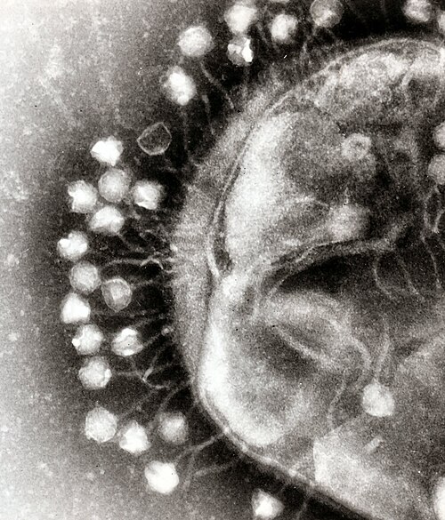

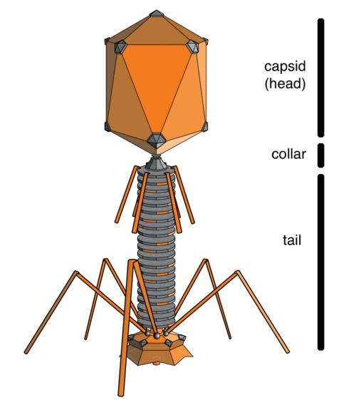


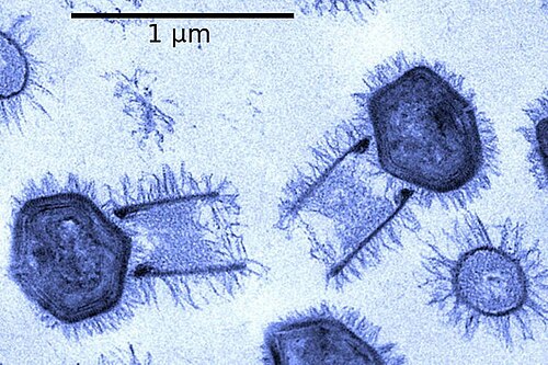
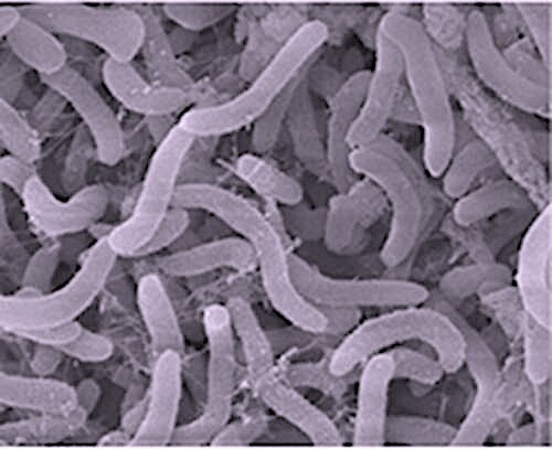


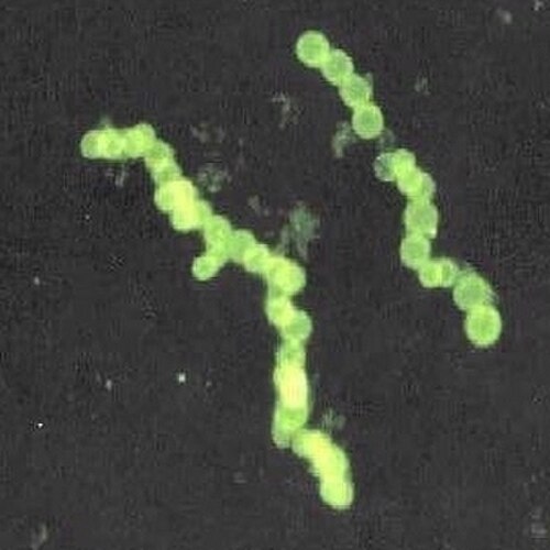 بكتيريا ثيومارغاريتا ناميبيينسيس البحرية ، أكبر بكتيريا معروفة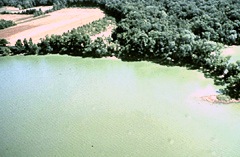
 تحتوي البلاستيدات الخضراء في الطحالب الزرقاء على طبقة من الببتيدوغليكان ، وهو دليل يشير إلى أصلها التكافلي الداخلي من البكتيريا الزرقاء . [ 72 ]
 تنمو بكتيريا مارينوموناس أركتيكا داخل الجليد البحري في القطب الشمالي في درجات حرارة تحت الصفر
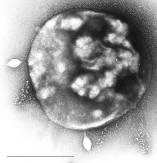 قد تكون الخلايا الأولية هي الأكثر وفرة بين العتائق البحرية
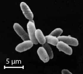 تُعرف البكتيريا المحبة للملح ، الموجودة في المياه المشبعة تقريبًا بالملح، الآن باسم العتائق.
 خلايا مسطحة مربعة الشكل من العتائق Haloquadratum walsbyi
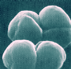 ميثانوسارسينا باركيري ، وهي عتائق بحرية تنتج الميثان
 تستطيع الكائنات المحبة للحرارة ، مثل فطر بيرولوبوس فوماري ، البقاء على قيد الحياة في درجات حرارة تزيد عن 100 درجة مئوية.
 من أين أتت الخلايا حقيقية النواة؟ – رحلة إلى عالم الميكروبات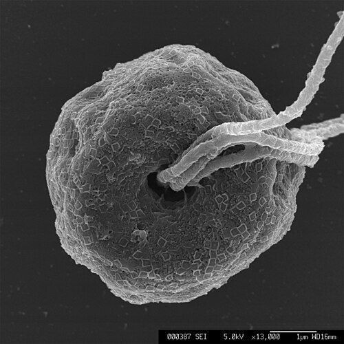

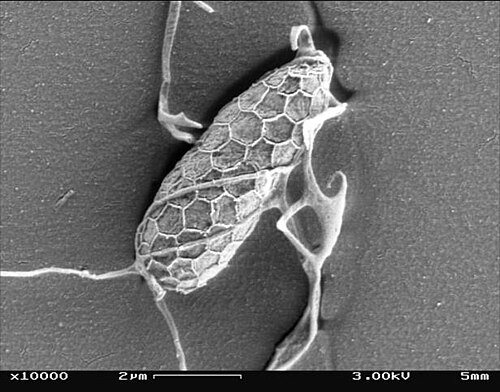

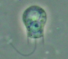
 تُعد الدياتومات مجموعة رئيسية من الطحالب، حيث تُنتج حوالي 20% من إنتاج الأكسجين العالمي. [ 93 ]
 هيكل دياتوم أحفوري يعود تاريخه إلى ما بين 32 و 40 مليون سنة
 طحلب وحيد الخلية، جيفيروكابسا أوشيانيكا
 اثنان من الدياتومات السوطية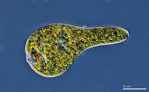
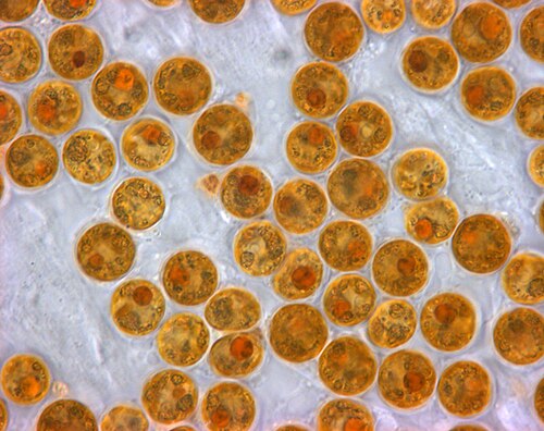

 هذا الهدبي يهضم البكتيريا الزرقاء . الفم الخلوي أو الفم يقع في أسفل اليمين.

 يبلغ طول Spiculosiphon oceana ، وهو كائن وحيد الخلية من فصيلة المنخربات ، شكله وأسلوب حياته يشبه الإسفنج ، 5 سم.
 يعيش زينوفيوفور ، وهو نوع آخر من المنخربات وحيدة الخلية، في المناطق السحيقة . وله صدفة عملاقة يصل قطرها إلى 20 سم. [ 97 ]
 عشب البحر العملاق ، وهو نوع من الطحالب البنية ، ليس نباتًا حقيقيًا، ولكنه متعدد الخلايا ويمكن أن ينمو إلى 50 مترًا




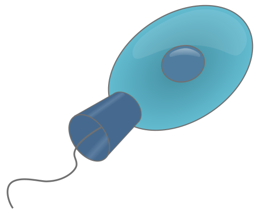
 سوط بكتيري يدور بواسطة محرك جزيئي في قاعدته
 حيوانات منوية سمك السلمون
 الهدبيات Oxytricha trifallax مع أهداب مرئية بوضوح
 أميبا مع دياتومات مبتلعة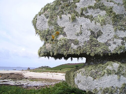

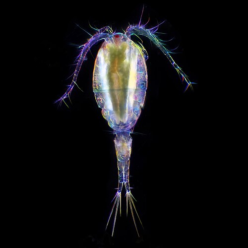 أكثر من 10000 نوع من الكائنات البحرية هي من مجدافيات الأرجل ، وهي قشريات صغيرة، وغالبًا ما تكون مجهرية.
 صورة فوتوغرافية بتقنية الحقل المظلم لحيوان يشبه الدودة ، يتراوح طوله بين 0.06 و3.0 ملم، ويعيش بين جزيئات الرواسب.
 يعيش نوع من الديدان المدرعة يُدعى Pliciloricus enigmaticus ، ويبلغ طوله حوالي 0.2 ملم، في الفراغات بين الحصى البحري.
 قد تبدو الدوارات ، التي يتراوح طولها عادةً بين 0.1 و0.5 ملم، مثل الطلائعيات، لكنها متعددة الخلايا وتنتمي إلى مملكة الحيوانات.
 تُعتبر الدببة المائية ( تارديغراد )، التي يبلغ طولها حوالي 0.5 ملم، من بين أكثر الحيوانات مرونة المعروفة

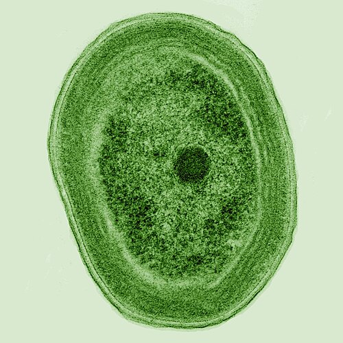
 الكلاميدوموناس غلوبوزا ، طحلب أخضر وحيد الخلية ذو سوطين يظهران بالكاد في أسفل اليسار
 الدياتوم المركزي
 الدياتومات
 سلاسل الطحالب الاستعمارية
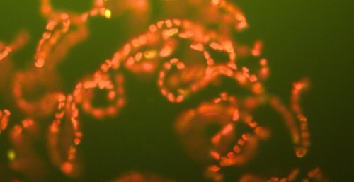
 العوالق النباتية هي أساس السلسلة الغذائية في المحيطات
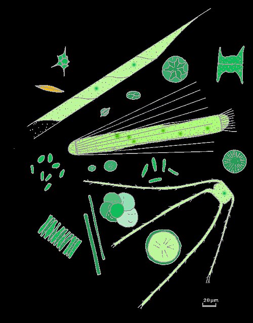 تأتي بأشكال وأحجام عديدة.
 العوالق النباتية المستعمرة
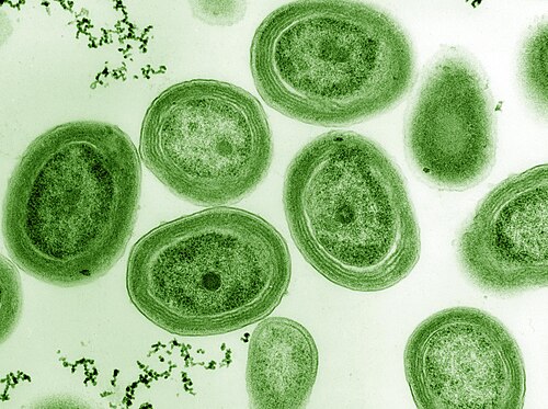 تُشكل البكتيريا الزرقاء Prochlorococcus جزءًا كبيرًا من الإنتاج الأولي للمحيطات
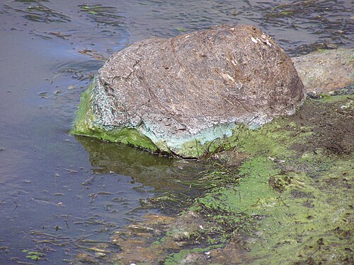 جرف الطحلب الأزرق الأخضر إلى صخرة في كاليفورنيا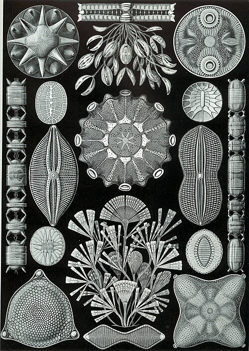

 تُعد الدياتومات من أكثر أنواع العوالق النباتية شيوعاً
 أغلفة الحماية الخاصة بها (الصدامات) مصنوعة من السيليكون
 تأتي بأشكال وأحجام عديدة

 Guinardia delicatula ، وهي دياتوم مسؤولة عن ازدهار الطحالب في بحر الشمال والقناة الإنجليزية [ 161 ].
 دياتوم أحفوري
 يوجد أكثر من 100,000 نوع من الدياتومات التي تمثل 50% من الإنتاج الأولي للمحيطات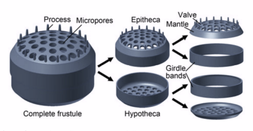
 تحتوي الكوكوليثوفورات على صفائح تسمى الكوكوليثات
 كوكوليثوس بيلاجيكوس ssp. braarudii
 Syracosphaera azureaplaneta ، سميت على اسم سلسلة الأفلام الوثائقية التي أنتجتها هيئة الإذاعة البريطانية The Blue Planet
 ازدهار طحالب إيميليانيا هوكسلي قبالة الساحل الجنوبي لإنجلترا
 أحفورة منقرضة

 تتغذى مجدافيات الأرجل على العوالق النباتية. وهذه المجدافية تحمل بيضاً.
 الهدبيات التينتينيدية فافيلا
 مثل الدياتومات، تأتي الشعاعيات بأشكال عديدة
 وكما هو الحال مع الدياتومات، فإن أصداف الشعاعيات تتكون عادة من السيليكات.
 لكنّ الشعاعيات الأكانثارية لها أصداف مصنوعة من بلورات كبريتات السترونتيوم.
 رسم تخطيطي مقطعي لصدفة شعاعية كروية
 Cladococcus abietinus


 مقطع يوضح حجرات ثقوب حلزونية
 يتدفق الإكتوبلازم الحبيبي الحي للأمونيا الدافئة لاصطياد الطعام
 مجموعة من المنخربات العوالقية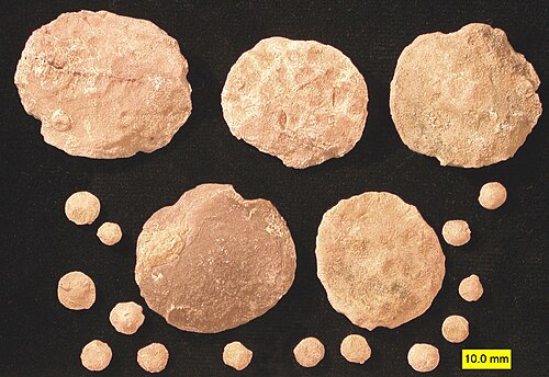
 الأميبا الخصية , Cyphoderia sp.
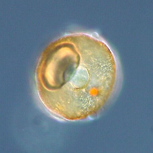 قشرة أو غلاف الأميبا ذات القشرة ، من نوع Arcella sp.
 أميبا غريبة المنشأ مغطاة بالدياتومات (من مجموعة بينارد للأميبا )
 الأميبا العارية، من نوع Chaos sp.
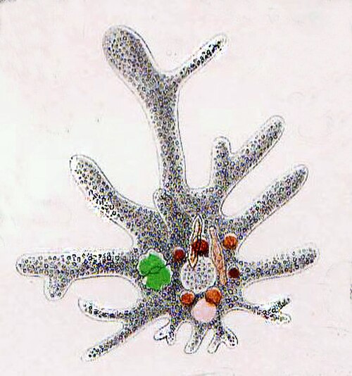 أميبا عارية تظهر فجوات غذائية ودياتوم مبتلع

 هولوفيرا أوفوم
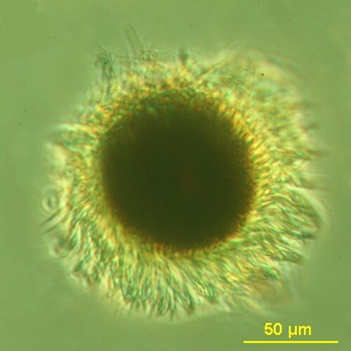 تنتج طحالب Mesodinium rubrum أزهارًا حمراء داكنة باستخدام البلاستيدات الخضراء المستعبدة من فرائسها من الطحالب [ 191 ].
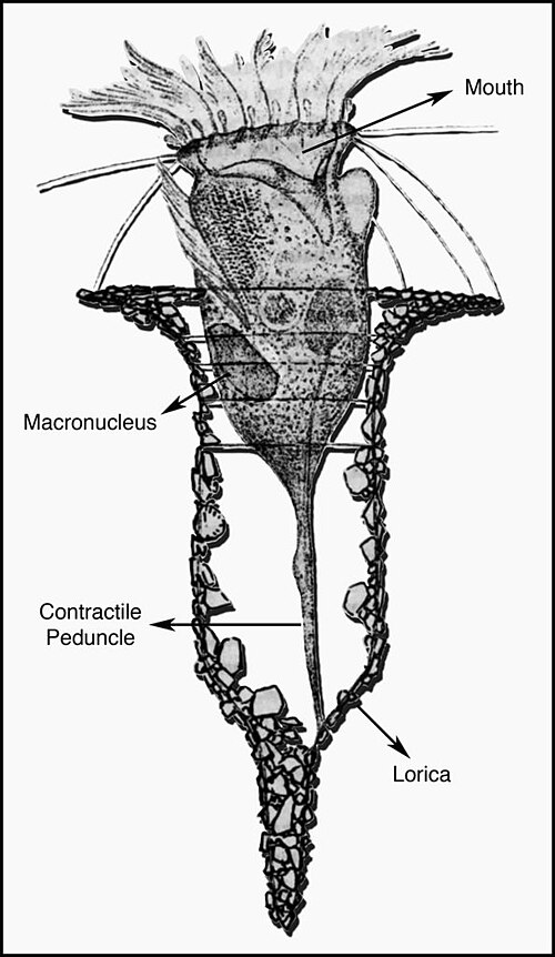


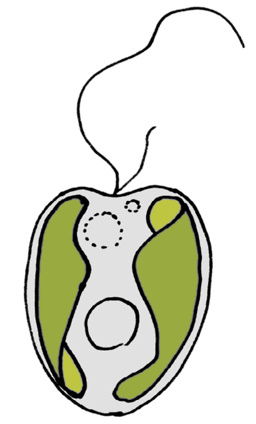
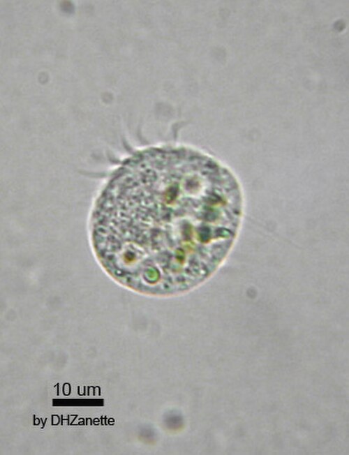


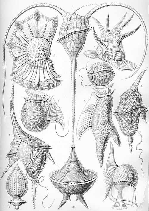
 Gyrodinium, one of the few naked dinoflagellates which lack armour
 The dinoflagellate Protoperidinium extrudes a large feeding veil to capture prey
 Nassellarian radiolarians can be in symbiosis with dinoflagellates
 The dinoflagellate Dinophysis acuta

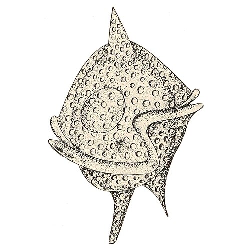

 Tripos muelleri is recognisable by its U-shaped horns
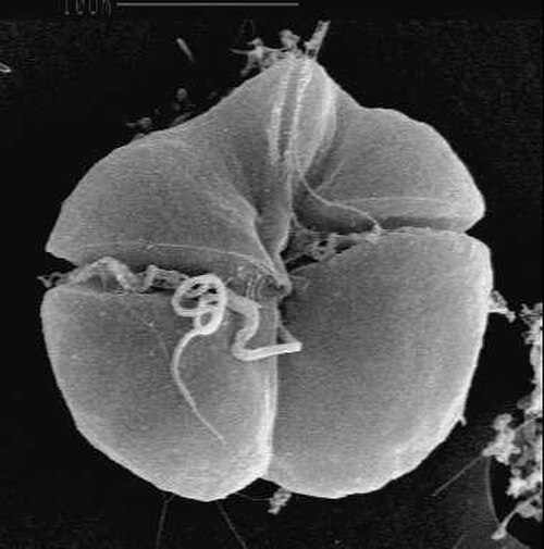 Karenia brevis produces red tides highly toxic to humans[214]
 Noctiluca scintillans, a bioluminescent dinoflagellate[215]

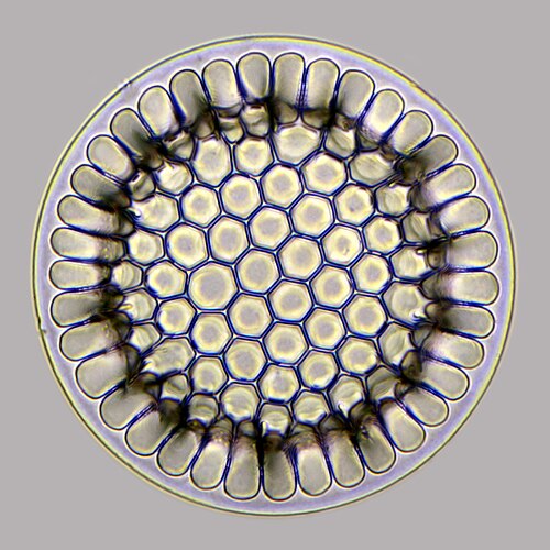


 Diatomaceous earth is a soft, siliceous, sedimentary rock made up of microfossils in the form of the frustules (shells) of single cell diatoms (click to magnify)
 Calcareous microfossils from marine sediment consisting mainly of star-shaped discoaster with a sprinkling of coccoliths
 Shells (tests), usually made of calcium carbonate, from a foraminiferal ooze on the deep ocean floor
 Illustration of a Globigerina ooze
 Marble can contain protist microfossils of foraminiferans, coccolithophores, calcareous nannoplankton and algae, ostracodes, pteropods, calpionellids and bryozoa[222]
 Opal can contain protist microfossils of diatoms, radiolarians, silicoflagellates and ebridians[222]


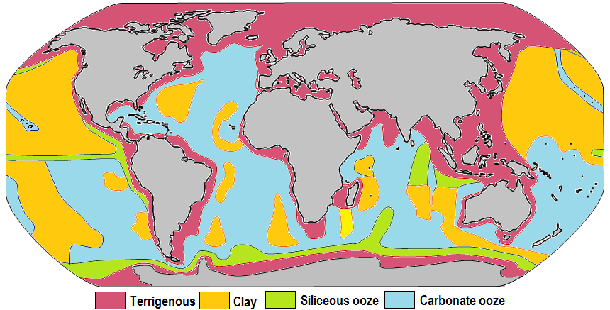 Distribution of sediment types on the seafloor Within each colored area, the type of material shown is what dominates, although other materials are also likely to be present.For further information, see here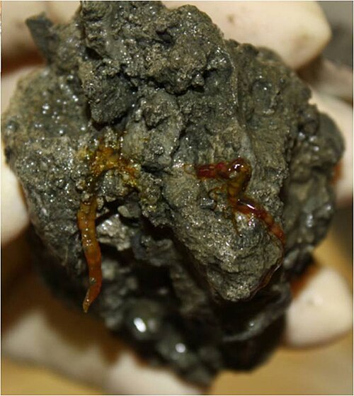
 Elphidium a widespread abundant genus of benthic forams
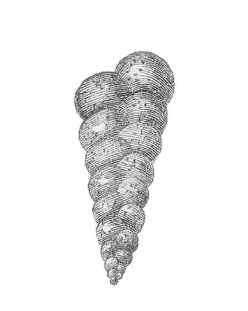 Heterohelix, an extinct genus of benthic forams

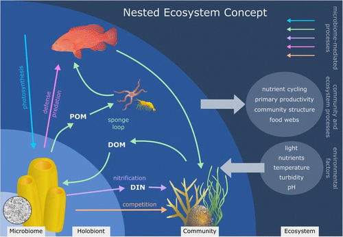


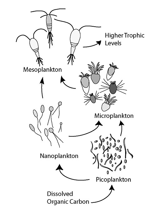


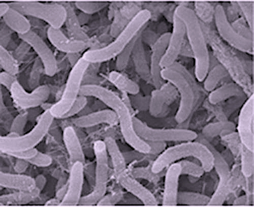 Pelagibacter ubique, the most abundant bacteria in the ocean, plays a major role in the global carbon cycle.
 Marine snow is the shower of organic particles that falls from upper waters to the deep ocean[248] It is a major exporter of carbon.


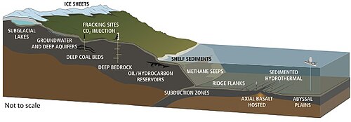


 Estimates of microbial species counts in the three domains of lifeBacteria are the oldest and most biodiverse group, followed by Archaea and Fungi (the most recent groups). In 1998, before awareness of the extent of microbial life had gotten underway, Robert M. May[274] estimated there were 3 million species of living organisms on the planet. But in 2016, Locey and Lennon[275] estimated the number of microorganism species could be as high as 1 trillion.[276]
 Microbial diversityComparative representation of the known and estimated (small box) and the yet unknown (large box) microbial diversity, which applies to both marine and terrestrial microorganisms. The text boxes refer to factors that adversely affect the knowledge of the microbial diversity that exists on the planet.[276]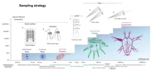




 Modified text was copied from this source, which is available under a Creative Commons Attribution 4.0 International License.
 This article incorporates text from this source, which is in the public domain.